. 2016 Sep 22;11(9):e0163216.
doi: 10.1371/journal.pone.0163216. eCollection 2016.

Production Conditions Affect the In Vitro Anti-Tumoral Effects of a High Concentration Multi-Strain Probiotic Preparation

Affiliations

Production Conditions Affect the In Vitro Anti-Tumoral Effects of a High Concentration Multi-Strain Probiotic Preparation

Benedetta Cinque et al. PLoS One. .

Erratum in

Expression of concern in

Abstract

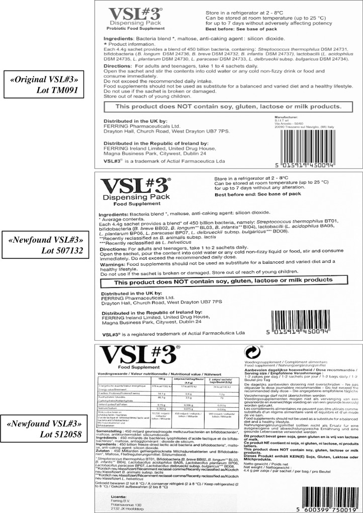
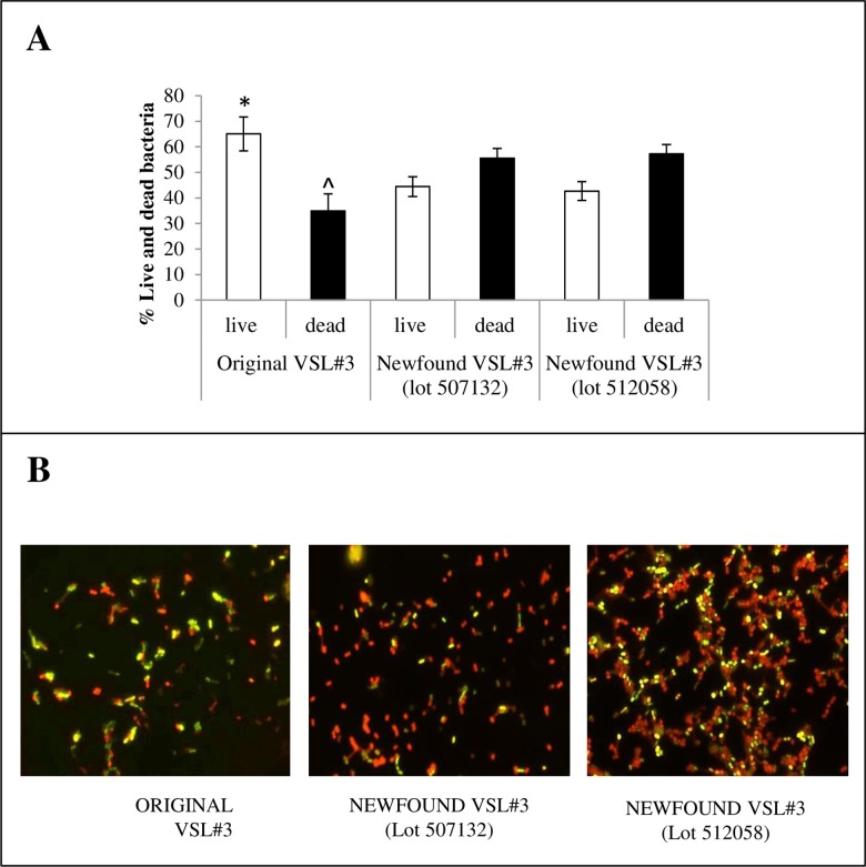
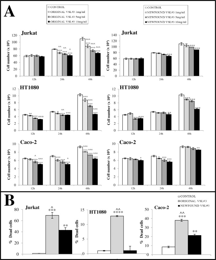
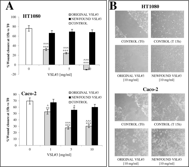
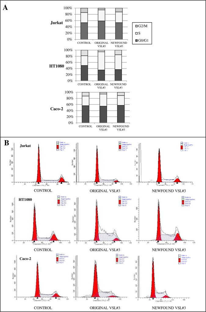
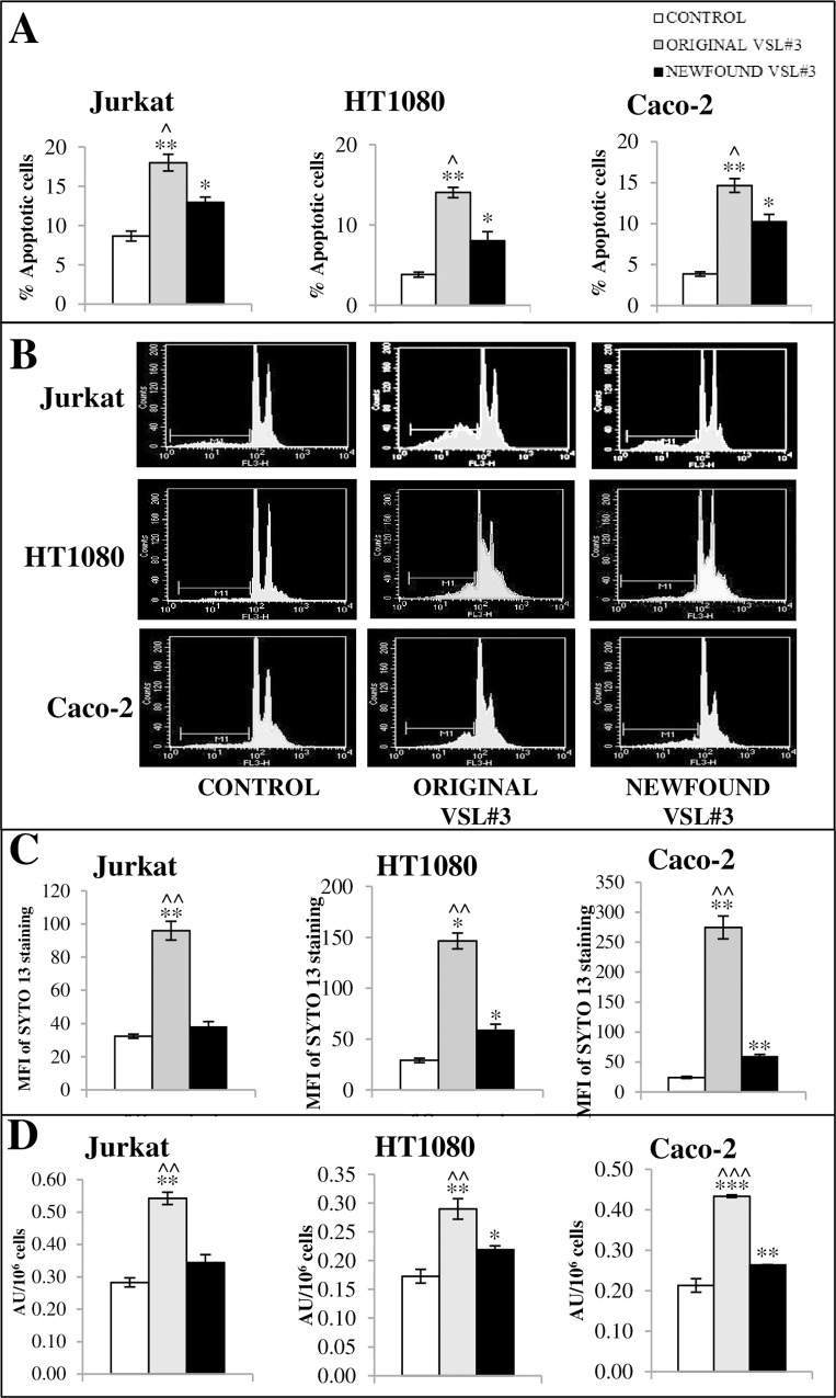
A careful selection of the probiotic agent, standardization of the dose and detailed characterization of the beneficial effects are essential when considering use of a probiotic for the dietary management of serious diseases. However, changes in the manufacturing processes, equipment or facilities can result in differences in the product itself due to the live nature of probiotics. The need to reconfirm safety and/or efficacy for any probiotic product made at a different factory is therefore mandatory. Recently, under the brand VSL#3®, a formulation produced by a manufacturer different from the previous one, has been commercialized in some European countries (the UK and Holland). VSL#3 is a high concentration multi-strain preparation which has been recognized by the main Gastroenterology Associations for the dietary management of pouchitis as well as ulcerative colitis. We have compared the "original" VSL#3 produced in USA with the "newfound" VSL#3 produced in Italy. According to our results, the "newfound" VSL#3 has 130-150% more "dead bacteria" compared to the "original" product, raising concerns for the well-known association between dead microbes with adverse effects. The abilities of bacterial lysates from the two formulations to influence in vitro viability and proliferation of different tumor cell lines also resulted different. The repair of previously scratched monolayers of various adherent tumor cell lines (i.e. HT1080, and Caco-2 cells) was inhibited more significantly by the "original" VSL#3 when compared to the "newfound" VSL#3. Tumor cell cycle profile, in particular cell cycle arrest and apoptotic death of the cancer cells, further confirms that the "original" VSL#3 has a better functional profile than the "newfound" VSL#3, at least in in vitro. Our data stress the importance of the production conditions for the "newfound" VSL#3 considering that this product is intended to be used for the dietary management of patients with very serious diseases, such as chronic inflammatory bowel diseases.


Conflict of interest statement

Dr. Van der Rest, the owner of BioVisible BV, has contributed to the present work as co-author. In particular, he provided his experience and expertise in the field of analysis related to the counts of bacteria. This does not alter our adherence to PLOS ONE policies on sharing data and materials.

Figures

Fig 1
Fig 1. “Original” and “newfound” VSL#3 formulations.
Fig 2
Fig 2. Live vs dead bacteria in “original” and “newfound” VSL#3 product.
Panel A: The percentages of live and dead cells in each VSL#3 formulations presented as mean ± SD of n. 10 determinations are shown. (Live cells *P<0.001 vs both lots of newfound formulations; Dead cells ^P<0.001 vs both lots of newfound formulations). Panel B: Representative microscopy images of bacteria stained with SYTO®13 and Propidium Iodide are shown. Stained bacterial suspensions were analyzed using a fluorescent microscope. Red colour is due to SYTO®13 green fluorescence that was quenched by that of PI (dead bacterial cells), while bright green-yellow colour was due to SYTO®13 staining when PI did not permeate across the cell membrane (live bacterial cells). Magnification 100x.
Fig 3
Fig 3
A) Comparison of the effects of “original” and “newfound” VSL#3 formulations on tumor cell growth. The cells were cultured for 12, 24 or 48 h in the presence or absence of probiotic lysates at 1, 5 and 10 mg/ml. The results from one representative from n. 3 independent experiments are presented as the mean values of duplicates ± SD. *P<0.05; **P<0.01; ***P<0.001 vs untreated control; ^P<0.05; ^^P<0.01; ^^^P<0.001 vs “newfound VSL#3” lysate treatment. All data shown are representative of 3 independent experiments. B) Comparison between the effects of bacterial lysates from each VSL#3 formulation on cell death % level on tumor cell lines. Dead cell number was assessed by Trypan blue dye inclusion after 48 h treatment in vitro. Percentages of dead cell number are presented as mean of duplicates. *P<0.05; **P<0.01; ***P<0.005; ****P<0.001 vs untreated control; ^P<0.05; ^^P<0.01 vs “newfound VSL#3” lysate treatment. All data shown are representative of 3 independent experiments.
Fig 4
Fig 4
A) Comparison between the effects of bacterial lysates on proliferation/migration ability of tumor cells assayed through in vitro “wound healing model”. The effect of bacterial lysates at 1, 5, and 10 mg/ml on the resurfacing of scratched monolayers was analyzed and compared to the relative control cells. The microscopy images acquired for each sample were analyzed with TScratch software to automatically quantify the percentage of wound closure. Data are presented as the mean of duplicate values ± SD) and are relative to % wound closure at 15 h from monolayer scratching. The results shown are representative of 3 independent experiments. *P<0.05, **P<0.01, and ***P<0.001 vs untreated control; ^P<0.05 and ^^^P<0.001 vs “newfound VSL#3” lysate treatment. B) Representative microscopy images of monolayers after treatment with “original” and “newfound VSL#3” formulations on adherent tumor cells at 15 h (T15) after scratching of monolayer. An image of scratched untreated monolayer for each used cell line at T0 is also shown. Images from one representative out of 3 independent experiments are shown.
Fig 5
Fig 5. Effect of bacterial lysates from “original” or “newfound VSL#3” products on tumor cell cycle distribution.
A) Histograms showing percent distribution of G0/G1, S, and G2/M cycle phase of tumor cells after 24 h treatment with probiotic lysates at 10 mg/ml. B) Representative cell cycles profiles of tumor cell lines after the same culture conditions above described.
Fig 6
Fig 6. Effect of bacterial lysates from “original” or “newfound VSL#3” products on tumor cell apoptosis level.
A) Data from cytofluorimetric analyses of apoptotic cells after treatment with bacterial lysates at 10 mg/ml for 48 hr expressed as mean % apoptotic cells ± SD. The data are relative to one representative out of n. 3 experiments in duplicate. *P<0.05, **P<0.01 vs untreated control; ^P<0.05 vs “newfound VSL#3” lysate treatment. B) The cytofluorimetric profiles from one representative from n. 3 experiments are shown. C) SYTO 13 staining of nucleic acid-containing microparticles generated in vitro from cell lines. Treated cells were incubated for 48 h with bacterial lysates, the media were collected and used for analysis of microparticles by flow cytometry. MFI, Mean Fluorescence Intensity. D) DNA fragmentation assay as measured by quantitation of cytosolic oligonucleosome-bound DNA, using an enzyme-linked immunosorbent assay (ELISA). Data from oligonucleosome assay after treatment with bacterial lysates at 10 mg/ml for 48 hr are expressed as Arbitrary Unit/106 cells (mean of duplicate values ± SD). The data are relative to one representative out of n. 3 experiments in duplicate. *P<0.05, **P<0.01, and ****P<0.001 vs untreated control; ^^P<0.01, and ^^^P<0.001 vs “original VSL#3” lysate treatment.

Similar articles

Cited by

References

    1. FAO/WHO (2002) Guidelines for the Evaluation of Probiotics in Food. Working Group Report 2002. Rome and Geneva: Food and Agricultural Organization of the United Nations/World Health Organization.
    1. Donelli G, Vuotto C, Mastromarino P. Phenotyping and genotyping are both essential to identify and classify a probiotic microorganism Microb Ecol Health Dis 2013, 24:20105 - PMC - PubMed
    1. Safety of Probiotics to Reduce Risk and Prevent or Treat Disease. AHRQ Publication No. 11-E007 April 2011. Agency for Healthcare Research and Quality, U.S. Department of Health and Human Services 540 Gaither Road Rockville, MD 20850 www.ahrq.gov
    1. Sanders ME, Klaenhammer TR, Ouwehand AC, et al. Effects of genetic, processing, or product formulation changes on efficacy and safety of probiotics. Annals of the New York Academy of Sciences 2014; 1309(1): 1–18. - PubMed
    1. Grześkowiak Ł, Isolauri E, Salminen S, Gueimonde M. Manufacturing process influences properties of probiotic bacteria. Br J Nutr. 2011. March;105(6):887–94. 10.1017/S0007114510004496 Epub 2010 Nov 9. - DOI - PubMed