. 2017 Feb 16;26(2):215-228.
doi: 10.3727/096368916X692951. Epub 2016 Sep 21.

A Nerve Conduit Containing a Vascular Bundle and Implanted With Bone Marrow Stromal Cells and Decellularized Allogenic Nerve Matrix

A Nerve Conduit Containing a Vascular Bundle and Implanted With Bone Marrow Stromal Cells and Decellularized Allogenic Nerve Matrix

Yukitoshi Kaizawa et al. Cell Transplant. .

Abstract

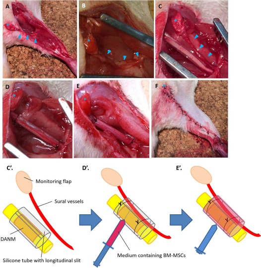
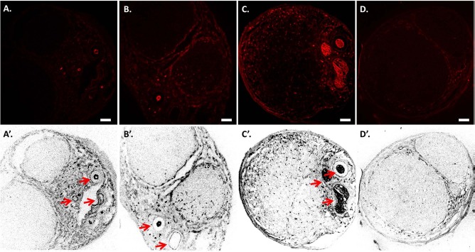
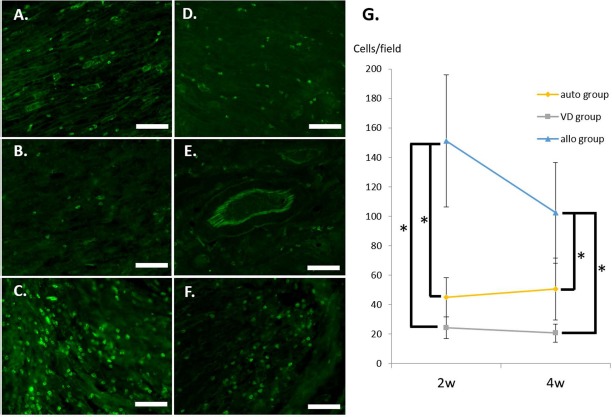
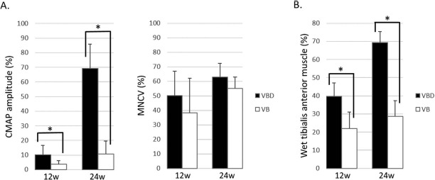
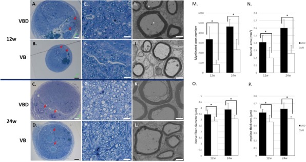
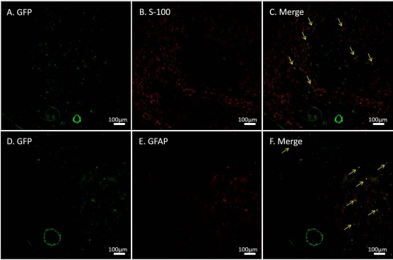
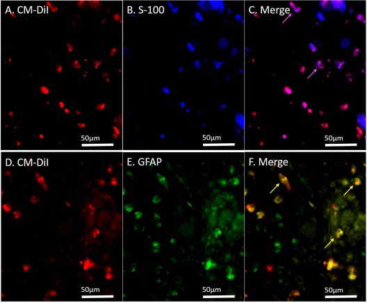
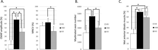
Cells, scaffolds, growth factors, and vascularity are essential for nerve regeneration. Previously, we reported that the insertion of a vascular bundle and the implantation of bone marrow-derived mesenchymal stem cells (BM-MSCs) into a nerve conduit promoted peripheral nerve regeneration. In this study, the efficacy of nerve conduits containing a vascular bundle, BM-MSCs, and thermally decellularized allogenic nerve matrix (DANM) was investigated using a rat sciatic nerve model with a 20-mm defect. Lewis rats were used as the sciatic nerve model and for the preparation of BM-MSCs, and Dark Agouti rats were used for the preparation of the DANM. The revascularization and the immunogenicity of the DANM were investigated histologically. The regeneration of nerves through nerve conduits containing vessels, BM-MSCs, and DANM (VBD group) was evaluated based on electrophysiological, morphometric, and reinnervated muscle weight measurements and compared with that of vessel-containing conduits that were implanted with BM-MSCs (VB group). The DANM that was implanted into vessel-containing tubes (VCTs) was revascularized by neovascular vessels that originated from the inserted vascular bundle 5-7 days after surgery. The number of CD8+ cells found in the DANM in the VCT was significantly smaller than that detected in the untreated allogenic nerve segment. The regenerated nerve in the VBD group was significantly superior to that in the VB group with regard to the amplitude of the compound muscle action potential detected in the pedal adductor muscle; the number, diameter, and myelin thickness of the myelinated axons; and the tibialis anterior muscle weight at 12 and 24 weeks. The additional implantation of the DANM into the BM-MSC-implanted VCT optimized the axonal regeneration through the conduit. Nerve conduits constructed with vascularity, cells, and scaffolds could be an effective strategy for the treatment of peripheral nerve injuries with significant segmental defects.


Figures

Figure 1
Figure 1
Surgical procedures used in the group with implantation of sural vessels, BM-MSCs, and a DANM (VBD group). (A, B) The sural flap was elevated, and a 15-mm nerve segment was resected. (C, C') The defect was bridged by a 20-mm decellularized allogenic nerve matrix (DANM) and subsequently inserted into a silicone tube through a slit in the tube. (D, D') A vascular pedicle with its monitoring flap was inserted into the silicon tube through the slit, and the slit was closed using 5-0 nylon suture. (E, E') α-MEM containing bone marrow-derived mesenchymal stem cells (BM-MSCs) was injected into the tube. (F) Postoperative appearance of the limb. The arrowheads indicate vascular bundles. The asterisk indicates the monitoring flap.
Figure 2
Figure 2
Revascularization of the decellularized allogenic nerve matrix (DANM). Immunohistochemistry for mouse monoclonal anti-rat endothelial cell cytoplasmic antigen (RECA-1) was performed on the transverse sections obtained from the midportion of the tube in the VD group at 5 days (A, A'), at 1 week (B, B'), and at 4 weeks (C, C') and in the D group at 4 weeks (D, D'). (A, A') A few RECA-1+ cells were detected only at the periphery of the DANM, and no RECA-1+ cells were observed inside the DANM at 5 days. (B, B') RECA-1+ cells were observed in the DANM at 1 week. The revascularized area was located mainly beside the inserted sural vessels. (C, C', D, and D') At 4 weeks, the RECA-1+ cells had spread diffusely in the DANM in the VD group (C, C'), whereas RECA-1+ cells were not found in the DANM in the D group (D, D'). The arrows indicate the inserted vascular bundle. Scale bars: 100 μm. VD group, the group with implantation of the sural vessels and a decellularized allogenic nerve matrix.
Figure 3
Figure 3
Immunogenicity of the grafts. Longitudinal sections of the distal part of the bridging materials were investigated in the auto group (A, D), the VD group (B, E), and the allo group (C, F) at 2 (A–C) and 4 (D–F) weeks using immunohistochemistry for CD8. The number of CD8+ cells was determined per field, and quantification is shown (G). Scale bars: 100 mm. ∗p < 0.01. VD group, the group with implantation of the sural vessels and a decellularized allogenic nerve matrix.
Figure 4
Figure 4
Electrophysiological studies and reinnervated muscle weight measurements (VBD group vs. VB group). The VBD group was significantly superior to the VB group regarding compound muscle action potential (CMAP) amplitude (A) and wet tibialis anterior muscle weight (B) at both 12 and 24 weeks. The data are presented as a percentage of the value recorded on the contralateral side. ∗p < 0.05. MNCV, motor nerve conduction velocity; VBD group, the group with implantation of the sural vessels, bone marrow-derived mesenchymal stem cells (BM-MSCs), and a decellularized allogenic nerve matrix (DANM); VB group, the group with the sural vessels and the BM-MSCs.
Figure 5
Figure 5
Histological and morphometric evaluations (VBD group vs. VB group). Transverse sections of the regenerated nerve 5 mm distal to the distal suture were observed under light microscopy (A–H) and transmission electron microscopy (TEM; I–L) at 12 (A, B, E, F, I, and J) and 24 (C, D, G, H, K, and L) weeks. Red blood cells filled the lumen of the inserted vessels (arrow). The number of myelinated axons (M) and the neural area (N) were larger in the VBD group compared with the VB group. Regenerated axons with a larger diameter (O) and thicker myelin sheaths (P) were observed in the VBD group compared with the VB group. Scale bars: 100 μm (A–D), 10 μm (E–H), and 2 μm (I–L). ∗p <0.05. VBD group, the group with implantation of the sural vessels, bone marrow-derived mesenchymal stem cells (BM-MSCs), and a decellularized allogenic nerve matrix (DANM); VB group, the group with the sural vessels and the BM-MSCs.
Figure 6
Figure 6
Immunohistochemistry for green fluorescent protein (GFP), S-100, and glial fibrillary acidic protein (GFAP). GFP+ cells were observed at 6 weeks after transplantation into conduits containing a vascular bundle and a decellularized allogenic nerve matrix (DANM). Some of the implanted cells were immunopositive for S-100 (A–C) and GFAP (D–F). The arrows indicate the merged cells.
Figure 7
Figure 7
Immunohistochemistry for S-100 and glial fibrillary acidic protein (GFAP) after transplantation of the chloromethylbenzamido dialkyl indocarbocyanine fluorescent dye (CM-DiI)-labeled bone marrow-derived mesenchymal stem cells (BM-MSCs). CM-DiI+ cells were detected in the decellularized allogenic nerve matrix (DANM) in the BM-MSC-seeded vessel-containing tube at 6 weeks after transplantation, and 31.8% and 28.8% of the CM-DiI+ cells were immunopositive for S-100 (A–C) and GFAP (D–F), respectively. The arrows indicate the merged cells.
Figure 8
Figure 8
Comparison of the regenerated nerve at 24 weeks (including the VD group, n = 8 in each group). The comparison among the VB, VBD, and VD groups at 24 weeks revealed that the VBD group was significantly superior not only to the VB group but also to the VD group regarding electrophysiological and morphometric measurements. The data obtained from electrophysiological studies (A) and reinnervated muscle weight measurements (C) are presented as a percentage of the value recorded on the contralateral side. The data were analyzed by one-way analysis of variance (ANOVA) followed by a Tukey–Kramer test. ∗p < 0.05 and #p = 0.06. CMAP, compound muscle action potential, MNCV, motor nerve conduction velocity; VB group, the group with the sural vessels and the bone marrow-derived mesenchymal stem cells (BM-MSCs); VBD group, the group with implantation of the sural vessels, the BM-MSCs, and a decellularized allogenic nerve matrix (DANM); VD group, the group with implantation of the sural vessels and a DANM.

References

    1. Meek MF, Coert JH. US Food and Drug Administration/Conformit Europe-approved absorbable nerve conduits for clinical repair of peripheral and cranial nerves. Ann Plast Surg. 2008; 60: 110–6. - PubMed
    1. Ray WZ, Mackinnon SE. Management of nerve gaps: Autografts, allografts, nerve transfers, and end-to-side neurorrhaphy. Exp Neurol. 2010; 223: 77–85. - PMC - PubMed
    1. Kakinoki R, Nishijima N, Ueba Y, Oka M, Yamamuro T. Relationship between axonal regeneration and vascularity in tubulation—An experimental study in rats. Neurosci Res. 1995; 23: 35–45. - PubMed
    1. Yamakawa T, Kakinoki R, Ikeguchi R, Nakayama K, Morimoto Y, Nakamura T. Nerve regeneration promoted in a tube with vascularity containing bone marrow-derived cells. Cell Transplant. 2007; 16: 811–22. - PubMed
    1. Kaizawa Y, Kakinoki R, Ikeguchi R, Ohta S, Noguchi T, Oda H, Matsuda S. Bridging a 30 mm defect in the canine ulnar nerve using vessel-containing conduits with implantation of bone marrow stromal cells. Microsurgery 2016; 36: 316–24. - PubMed