. 2016 Sep 22;11(9):e0163174.
doi: 10.1371/journal.pone.0163174. eCollection 2016.

The High-Risk Human Papillomavirus E6 Oncogene Exacerbates the Negative Effect of Tryptophan Starvation on the Development of Chlamydia trachomatis

Affiliations

The High-Risk Human Papillomavirus E6 Oncogene Exacerbates the Negative Effect of Tryptophan Starvation on the Development of Chlamydia trachomatis

Shardulendra P Sherchand et al. PLoS One. .

Abstract

Chlamydia trachomatis is an obligate intracellular pathogen that requires specific essential nutrients from the host cell, one of which is the amino acid tryptophan. In this context interferon gamma (IFNγ) is the major host protective cytokine against chlamydial infections because it induces the expression of the host enzyme, indoleamine 2,3-dioxygenase 1, that degrades tryptophan, thereby restricting bacterial replication. The mechanism by which IFNγ acts has been dissected in vitro using epithelial cell-lines such as HeLa, HEp-2, or the primary-like endocervical cell-line A2EN. All these cell-lines express the high-risk human papillomavirus oncogenes E6 & E7. While screening cell-lines to identify those suitable for C. trachomatis co-infections with other genital pathogens, we unexpectedly found that tryptophan starvation did not completely block chlamydial development in cell-lines that were HR-HPV negative, such as C33A and 293. Therefore, we tested the hypothesis that HR-HPV oncogenes modulate the effect of tryptophan starvation on chlamydial development by comparing chlamydial development in HeLa and C33A cell-lines that were both derived from cervical carcinomas. Our results indicate that during tryptophan depletion, unlike HeLa, C33A cells generate sufficient intracellular tryptophan via proteasomal activity to permit C. trachomatis replication. By generating stable derivatives of C33A that expressed HPV16 E6, E7 or E6 & E7, we found that E6 expression alone was sufficient to convert C33A cells to behave like HeLa during tryptophan starvation. The reduced tryptophan levels in HeLa cells have a biological consequence; akin to the previously described effect of IFNγ, tryptophan starvation protects C. trachomatis from clearance by doxycycline in HeLa but not C33A cells. Curiously, when compared to the known Homo sapiens proteome, the representation of tryptophan in the HR-HPV E6 & E6AP degradome is substantially lower, possibly providing a mechanism that underlies the lowered intracellular free tryptophan levels in E6-expressing cells during starvation.


Conflict of interest statement

The authors have declared that no competing interests exist.

Figures

Fig 1
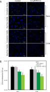
Fig 1. Cell-line dependent differences in the effect of tryptophan depletion on C. trachomatis development.
Cells were infected with C. trachomatis at an m.o.i of 5 as described in the methods. After infection cells were maintained in complete media or tryptophan-free media. After 42 h.p.i. cells are either stained by immunofluorescence for chlamydial LPS using a FITC-conjugated antibody, or harvested to quantify IFU recovery by infection of HeLa cells. A) Chlamydial inclusions (green) formed in HeLa, A2EN, C33A and 293 cells 42 h.p.i. Cells were counterstained with Hoechst dye. Scale bar indicates 20 μm. B) IFU/mL recovered after 42 h.p.i. in HeLa, A2EN, C33A and 293 cells as evaluated by infection of HeLa cells. The bars represents mean and standard deviation from three independent experiments (** indicates P < 0.01 by the Wilcoxon rank sum test).
Fig 2
Fig 2. Temporal evaluation of eIF2α phosphorylation in HeLa and C33A grown in tryptophan-free media.
HeLa and C33A cells were plated in complete media, which was replaced with tryptophan-free media 24 hours post-plating. Cells were harvested every 6 hours and used to make extracts that were evaluated by immunoblot using antibodies against eIF2α or eIF2α phosphorylated on serine 51 (p-eIF2α). Immunoblots against β-actin were performed as a loading control. Similar results were obtained from three independent experiments.
Fig 3
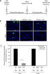
Fig 3. The efficacy of doxycycline treatment against C. trachomatis during tryptophan starvation is cell-line dependent.
A) Schematic depiction of the experimental protocol. HeLa or C33A cells were infected with C. trachomatis. Post-infection, cells were maintained in complete media (CM), complete media with 1 μg/mL doxycycline (CM/Dox), or tryptophan-free media with 1 μg/mL doxycycline (TF/Dox). Infected cells were exposed to doxycycline for 36 hours, following which the media was replaced with complete media. After an additional 36 hours of growth in complete media, infected cells were evaluated for inclusion formation and IFU recovery. B) Immunofluorescence for chlamydial LPS to detect inclusions in infected HeLa and C33A cells under the three conditions described in “A”, i.e. CM + CM, CM/Dox + CM, and TF/Dox + CM. Cells were counterstained with Hoechst dye, and a scale bar of 20 μm is indicated. C) IFU/mL recovered under the three conditions indicated in “A” as a percent of the IFU/mL recovered under the CM + CM condition. IFU recovery was quantified by infection of HeLa cells. The bars represent the mean and standard deviation from three independent experiments. Significantly higher IFUs were recovered when infected HeLa cells were initially grown in the TF/Dox condition relative to the CM/Dox condition (* indicates p < 0.05 by the Wilcoxon rank sum test).
Fig 4
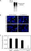
Fig 4. Proteasome activity is essential to support chlamydial development in C33A cells during tryptophan starvation.
Immediately after infection with C. trachomatis, C33A cells were grown under four conditions: Complete media + vehicle control; Complete Media + 0.3 μM MG132; Trp-free media + vehicle control; and Trp-free media with 0.3 μM MG132. After 42 hours of growth under these conditions, inclusion development and IFU recovery was evaluated. A) Immunoblot evaluating the accumulation of polyubiquitinated proteins in C33A cells 24 hours post-exposure to the vehicle control or 0.3 μM MG132. The primary antibody used a rabbit polyclonal anti-ubiquitin antibody. B) Immunofluorescence using a FITC-conjugated anti-chlamydial LPS antibody was used to evaluate inclusion formation under the four conditions described above. Cells were counterstained with Hoechst dye. C) IFU/mL recovered 42 h.p.i. from cells grown under the four conditions described above. IFU recovery was quantified by infection of HeLa cells. The addition of 0.3 μM MG132 dramatically reduced IFU/mL released from C33A cells grown in Trp-Free Media (** indicates p < 0.001 by the Wilcoxon rank sum test). The bars represent the mean and standard deviation from three independent experiments.
Fig 5
Fig 5. High levels of IFNγ partially restores C. trachomatis development in HeLa cells.
After infection with C. trachomatis, HeLa cells were grown in DMEM containing 4 mg/L of tryptophan, and treated with 0, 300, 1500 U/mL of IFNγ, or I500 U/mL IFNγ + 0.3 μM MG132 for 42 hours, following which inclusion formation and IFU recovery were evaluated. A) Immunofluorescence using a FITC-conjugated anti-chlamydial LPS antibody was used to evaluate inclusion formation under the four conditions described above. Cells were counterstained with Hoechst dye. B) IFU/mL recovered at 42 h.p.i. from cells grown under the four conditions described above. IFU recovery was quantified by infection of HeLa cells. As reported previously [19], the addition of 300 U/mL IFNγ resulted in a statistically significant 2-log decrease in IFU recovery. Addition of 1500 U/mL IFNγ resulted in a statistically significant ~1-log increase in the IFU recovered relative to the addition of 300 U/mL IFNγ. IFU recovery was addition of 1500 U/mL IFNγ was significantly reduced by the concurrent administration of 0.3 μM MG132. The data represent three independent experiments (** indicates P < 0.001 by the Wilcoxon rank sum test).
Fig 6
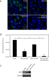
Fig 6. Expression of the HPV16 E6 oncogene in C33A cells accentuates the effect of tryptophan starvation on C. trachomatis development.
Stable C33A-derivative cell-lines were constructed by transducing C33A cells with a control retroviral vector (pLXSN), or pLXSN derivatives expressing the HPV16 E6, E6 & E7, or E7 oncogenes. Stable cell-lines were selected using G418 as described in the material and method section. A) Cell-lines were grown in tryptophan-free media for 12 hours after which immunoblots were used to query the phosphorylation status of eIF2α. Total eIF2α was used as a loading control. B) IFU/mL recovered at 42 h.p.i. after infected cells were grown in complete media or tryptophan free-media, as evaluated by infection of HeLa cells. As anticipated, growth in tryptophan-free media reduced IFU/mL recovered from all four cell-lines. IFU/mL recovery from C33A/E6 and C33A/E6+E7 cell-lines during tryptophan starvation were significantly lower than the IFU/mL recovered from the control C33A/pLXSN cell-line. The data represent three independent experiments (** indicates P < 0.01 by the Wilcoxon rank sum test).
Fig 7
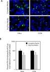
Fig 7. Arginine starvation impacts C. trachomatis development in HeLa and C33A cells to an equivalent extent.
C. trachomatis infected HeLa and C33A cells were grown in Complete Media and arginine-free (Arg-Free) media for 42 h.p.i. at which inclusion development and IFU recovery were evaluated. A) Immunofluorescence using a FITC-conjugated anti-chlamydial LPS antibody was used to evaluate inclusion formation in HeLa and C33A cells grown in Complete Media or Arg-Free Media. Cells were counterstained with Hoechst dye. B) IFU/mL recovered at 42 h.p.i. after infected cells were grown in complete media or arginine free-media, as evaluated by infection of HeLa cells. Although the IFU/mL recovered under arginine starvation conditions was lower than that observed in complete media, the decrease was not observed to be statistically significant for either HeLa or C33A cells (P > 0.05 by the Wilcoxon rank sum test).
Fig 8
Fig 8. Proteasome inhibition equivalently affects C. trachomatis growth in HeLa and C33A during arginine starvation.
C. trachomatis infected HeLa and C33A cells were grown in complete media (CM) or arginine free media (Arg-Free) with a vehicle control (CM, Arg-Free) or in the presence of 0.3 μM MG132 (CM + MG132, Arg-Free + MG132) for 42 h.p.i. At this time, cells were either stained to observe inclusion size or harvested to quantify IFU released. A) Immunofluorescence using a FITC-conjugated anti-chlamydial LPS antibody was used to evaluate inclusion formation in HeLa and C33A cells grown in CM, CM + MG132, Arg-Free, or Arg-Free + MG132. Cells were counterstained with Hoechst dye. B) IFU/mL recovered at 42 h.p.i. after infected cells were grown under the conditions used in “A”. IFU/mL was quantified by infection of HeLa cells. Although the IFU/mL recovered during arginine starvation was lower than that observed in complete media, the decrease was not observed to be statistically significant for either HeLa or C33A cells (P > 0.05 by the Wilcoxon rank sum test). The addition of MG132 did not affect IFU release for cells grown in CM. A statistically significant decrease (P < 0.05) was observed for cells grown in Arg-Free media.

References

    1. Wolf K, Fischer E, Hackstadt T. Ultrastructural analysis of developmental events in Chlamydia pneumoniae-infected cells. Infect Immun. 2000;68(4):2379–85. Epub 2000/03/18. - PMC - PubMed
    1. Moulder JW. Interaction of chlamydiae and host cells in vitro. Microbiol Rev. 1991;55(1):143–90. Epub 1991/03/01. - PMC - PubMed
    1. Brunham RC, Rey-Ladino J. Immunology of Chlamydia infection: implications for a Chlamydia trachomatis vaccine. Nat Rev Immunol. 2005;5(2):149–61. Epub 2005/02/03. 10.1038/nri1551 . - DOI - PubMed
    1. Stephens RS, Kalman S, Lammel C, Fan J, Marathe R, Aravind L, et al. Genome sequence of an obligate intracellular pathogen of humans: Chlamydia trachomatis. Science. 1998;282(5389):754–9. Epub 1998/10/23. . - PubMed
    1. Abdelrahman YM, Belland RJ. The chlamydial developmental cycle. FEMS Microbiol Rev. 2005;29(5):949–59. Epub 2005/07/27. 10.1016/j.femsre.2005.03.002 . - DOI - PubMed