. 2016 Dec;14(12):1266-1276.
doi: 10.1158/1541-7786.MCR-16-0233. Epub 2016 Sep 22.

MUC1-C Represses the Crumbs Complex Polarity Factor CRB3 and Downregulates the Hippo Pathway

Affiliations

MUC1-C Represses the Crumbs Complex Polarity Factor CRB3 and Downregulates the Hippo Pathway

Maroof Alam et al. Mol Cancer Res. 2016 Dec.

Abstract

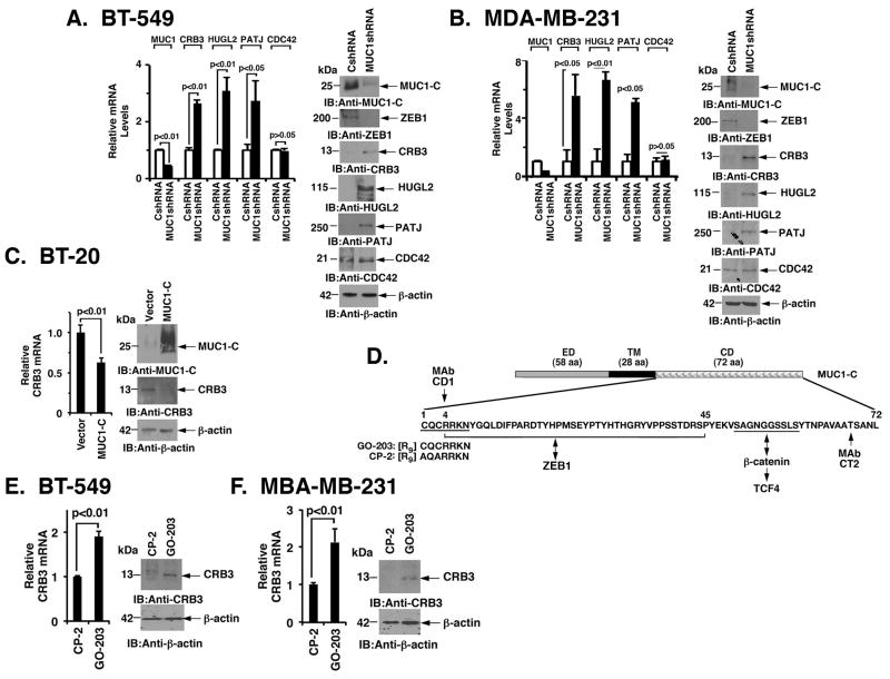
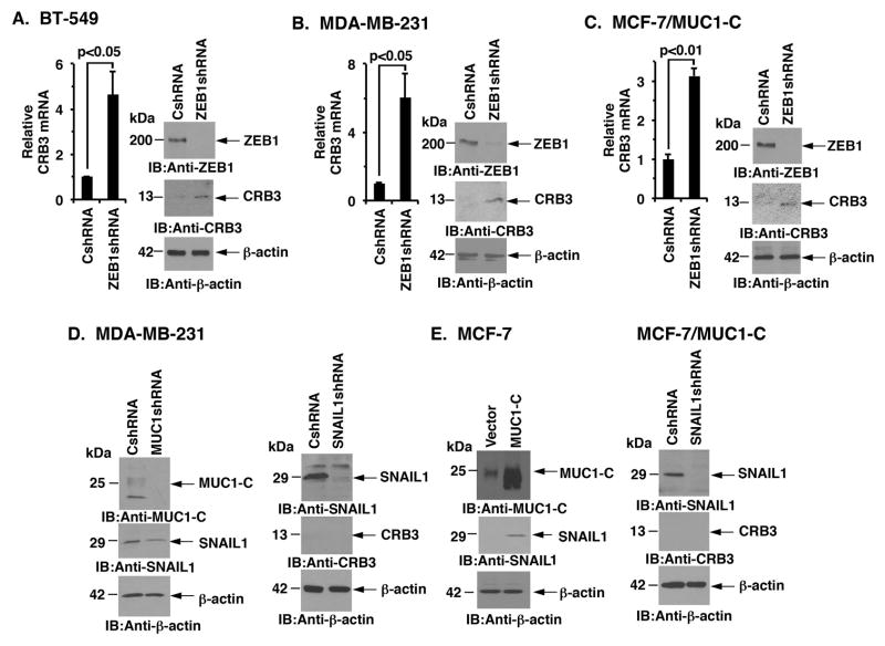
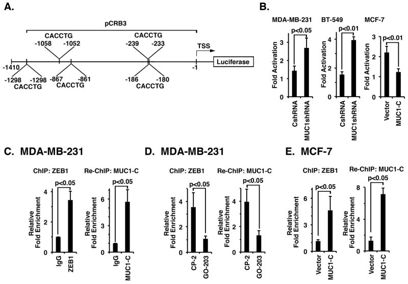
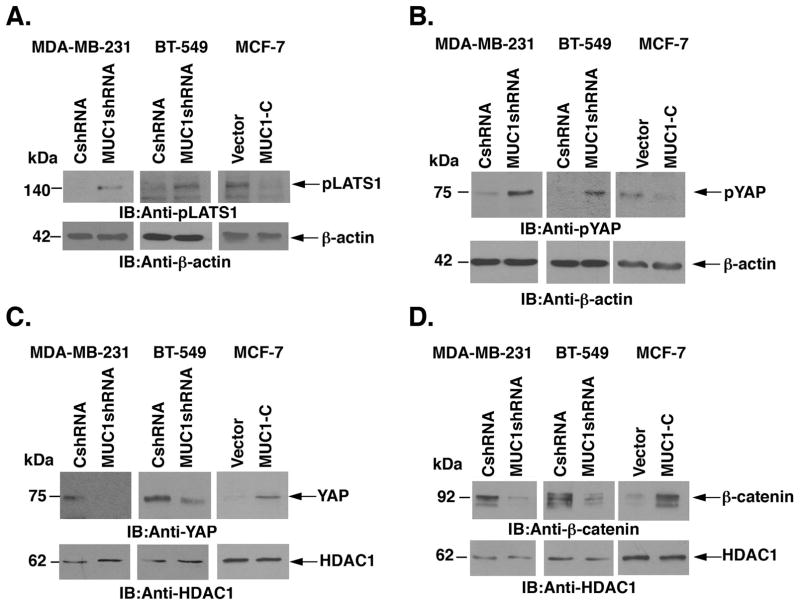
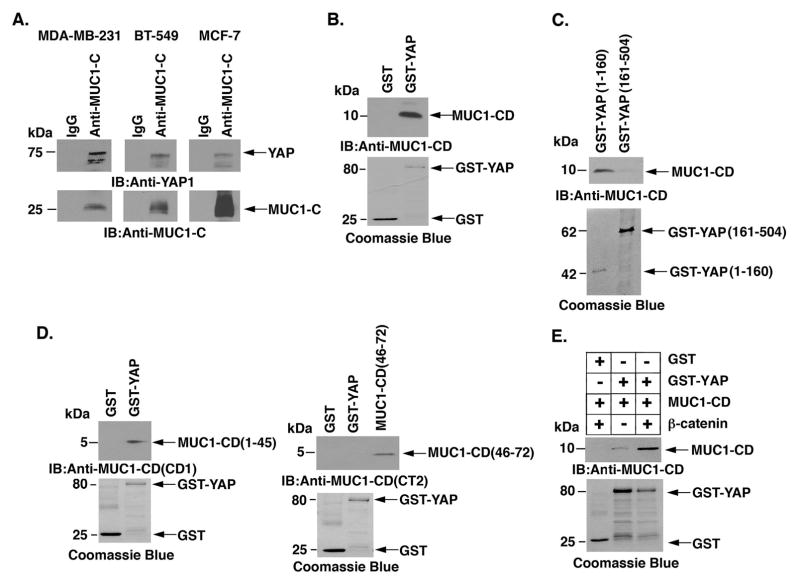
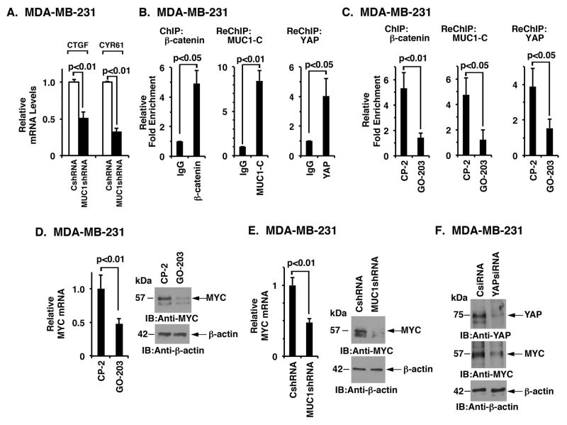
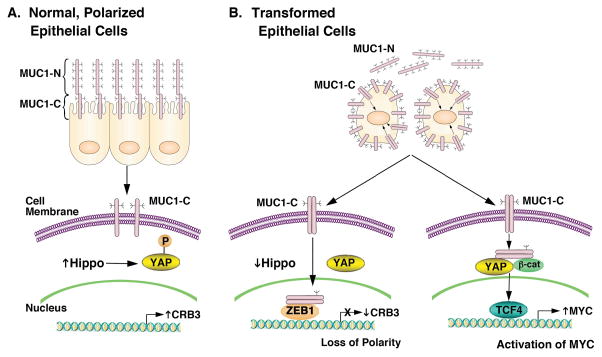
Apical-basal polarity and epithelial integrity are maintained in part by the Crumbs (CRB) complex. The C--terminal subunit of MUC1 (MUC1-C) is a transmembrane protein that is expressed at the apical border of normal epithelial cells and aberrantly at high levels over the entire surface of their transformed counterparts. However, it is not known whether MUC1-C contributes to this loss of polarity that is characteristic of carcinoma cells. Here it is demonstrated that MUC1-C downregulates expression of the Crumbs complex CRB3 protein in triple-negative breast cancer (TNBC) cells. MUC1-C associates with ZEB1 on the CRB3 promoter and represses CRB3 transcription. Notably, CRB3 activates the core kinase cassette of the Hippo pathway, which includes LATS1 and LATS2. In this context, targeting MUC1-C was associated with increased phosphorylation of LATS1, consistent with activation of the Hippo pathway, which is critical for regulating cell contact, tissue repair, proliferation, and apoptosis. Also shown is that MUC1-C--mediated suppression of CRB3 and the Hippo pathway is associated with dephosphorylation and activation of the oncogenic YAP protein. In turn, MUC1-C interacts with YAP, promotes formation of YAP/β-catenin complexes, and induces the WNT target gene MYC. These data support a previously unrecognized pathway in which targeting MUC1-C in TNBC cells (i) induces CRB3 expression, (ii) activates the CRB3-driven Hippo pathway, (iii) inactivates YAP, and thereby (iv) suppresses YAP/β-catenin-mediated induction of MYC expression.

Implications: These findings demonstrate a previously unrecognized role for the MUC1-C oncoprotein in the regulation of polarity and the Hippo pathway in breast cancer. Mol Cancer Res; 14(12); 1266-76. ©2016 AACR.


Conflict of interest statement

Potential Conflict of Interest: The authors declare competing financial interests: D.K. holds equity in Genus Oncology and is a consultant to the company. The other authors disclosed no potential conflicts of interest.

Figures

Figure 1
Figure 1. MUC1-C downregulates CRB3 expression
A, BT-549 cells infected with lentiviruses to stably express a control scrambled shRNA (CshRNA) or a MUC1shRNA were analyzed for MUC1, CRB3, HUGL2, PATJ and CDC42 mRNA levels by qRT-PCR (left). The results are expressed as relative mRNA levels (mean±SD of three determinations) as compared with that obtained for BT-549/CshRNA cells (assigned a value of 1). Lysates from the BT-549/CshRNA and BT-549/MUC1shRNA cells were immunoblotted with the indicated antibodies (right). B, MDA-MB-231/CshRNA and MDA-MB-231/MUC1shRNA cells were analyzed for MUC1, CRB3, HUGL2, PATJ and CDC42 mRNA levels by qRT-PCR (left). The results are expressed as relative mRNA levels (mean±SD of three determinations) as compared with that obtained for MDA-MB-231/CshRNA cells (assigned a value of 1). Lysates from the MDA-MB-231/CshRNA and MDA-MB-231/MUC1shRNA cells were immunoblotted with the indicated antibodies (right). C, BT-20 cells stably expressing an empty vector or MUC1-C were analyzed for CRB3 mRNA levels by qRT-PCR (left). The results are expressed as relative CRB3 mRNA levels (mean±SD of three determinations) as compared to that obtained for BT-20/vector cells (assigned a value of 1). Lysates from the BT-20/vector and BT-20/MUC1-C cells were immunoblotted with the indicated antibodies (right). D, Schematic showing the MUC1-C subunit and the amino acid (aa) sequence of the cytoplasmic domain (CD). ED, extracellular domain; TM, transmembrane domain. The CQC motif is necessary and sufficient for MUC1-C homodimerization. D-amino acid sequences are shown for GO-203 and CP-2. E, BT-549 cells were treated with 5 μM GO-203 or CP-2 each day for 3 days and then analyzed for CRB3 mRNA levels by qRT-PCR (left). Lysates from BT-549 cells treated as above were immunoblotted with the indicated antibodies (right). F, MDA-MB-231 cells were treated with 5 μM GO-203 or CP-2 each day for 3 days and then analyzed for CRB3 mRNA levels by qRT-PCR (left), while lysates from similarly treated cells were immunoblotted with the indicated antibodies (right).
Figure 2
Figure 2. MUC1-C suppresses CRB3 expression by a ZEB1-dependent mechanism
A, BT-549 cells infected with lentiviruses to stably express a control scrambled shRNA (CshRNA) or a ZEB1shRNA were analyzed for CRB3 mRNA levels by qRT-PCR (left). The results are expressed as relative CRB3 mRNA levels (mean±SD of three determinations) as compared with that obtained for BT-549/CshRNA cells (assigned a value of 1). Lysates from the BT-549/CshRNA and BT-549/ZEB1shRNA cells were immunoblotted with the indicated antibodies (right). B, MDA-MB-231 cells stably expressing a control scrambled shRNA (CshRNA) or a ZEB1 shRNA were analyzed for CRB3 mRNA levels by qRT-PCR (left). The results are expressed as relative CRB3 mRNA levels (mean±SD of three determinations) as compared with that obtained for MDA-MB-231/CshRNA cells (assigned a value of 1). Lysates from the MDA-MB-231/CshRNA and MDA-MB-231/ZEB1shRNA cells were immunoblotted with the indicated antibodies (right). C, MCF-7/MUC1-C cells stably expressing a control scrambled shRNA (CshRNA) or a ZEB1 shRNA were analyzed for CRB3 mRNA levels by qRT-PCR (left). Lysates from MCF-7/MUC1-C/CshRNA and MCF-7/MUC1-C/ZEB1shRNA cells were immunoblotted with the indicated antibodies (right). D, Lysates from MDA-MB-231/CshRNA and MDA-MB-231/MUC1shRNA (left) and from MDA-MB-231/CshRNA and MDA-MB-231/SNAIL1shRNA (right) cells were immunoblotted with the indicated antibodies. E, Lysates from MCF-7/vector and MCF-7/MUC1-C cells (left) were immunoblotted with the indicated antibodies. Lysates from MCF-7/MUC1-C cells infected with lentiviruses to stably express a control scrambled shRNA (CshRNA) or a SNAIL1shRNA were immunoblotted with the indicated antibodies (right).
Figure 3
Figure 3. MUC1-C represses CRB3 promoter activation by a ZEB1-dependent mechanism
A, Schema of CRB3 luciferase reporter plasmid. B, MDA-MB-231/CshRNA and MDA-MB-231/MUC1shRNA cells (left), BT-549/CshRNA and BT-549/MUC1shRNA cells (middle) and MCF-7/Vector and MCF-7/MUC1-C cells (right) were transfected with the empty Luc vector or pCRB3-Luc. Cells were also transfected with the SV-40-Renilla-Luc plasmid as an internal control. Luciferase activity was measured at 48 h following transfection. The results are expressed as relative luciferase activity (mean±SD of three determinations) compared with that obtained from cells transfected with the empty Luc vector (assigned a value of 1). C (left), Soluble chromatin from the indicated MDA-MB-231 cells was precipitated with anti-ZEB1 or a control IgG. The final DNA precipitates were amplified by qPCR with pairs of primers for the ZEB1 binding region in CRB3 promoter region. Results are expressed as the relative fold enrichment (mean±SD of three determinations) compared with that obtained for the IgG control (assigned a value of 1). For re-ChIP analysis, soluble chromatin from MDA-MB-231 cells (right) was first precipitated with anti-ZEB1, then released and reimmunoprecipitated with anti-MUC1-C. The results are expressed as the relative fold enrichment (mean±SD of three determinations) compared with that obtained with the IgG control (assigned a value of 1). D, MDA-MB-231 cells were treated with 5 μM GO-203 or CP-2 each day for 3 days. Soluble chromatin was precipitated with anti-ZEB1 or a control IgG (left). For re-ChIP analysis, complexes were released and re-immunoprecipitated with anti-MUC1-C (right). Results are expressed as the relative fold enrichment (mean±SD of three determinations) compared with that obtained for the IgG control (assigned a value of 1). E, Soluble chromatin from MCF-7/vector and MCF-7/MUC1-C cells was precipitated with anti-ZEB1 or a control IgG (left). For re-ChIP analysis, complexes were released and re-immunoprecipitated with anti-MUC1-C (right). Results are expressed as the relative fold enrichment (mean±SD of three determinations) compared with that obtained for the IgG control (assigned a value of 1).
Figure 4
Figure 4. MUC1-C-mediated CRB3 repression increases nuclear accumulation of YAP
A, Lysates from MDA-MB-231/CshRNA and MDA-MB-231/MUC1shRNA cells (left), BT-549/CshRNA and bt-549/MUC1shRNA (middle) and MCF-7/Vector and MCF-7/MUC1-C (right) were immunoblotted with the indicated antibodies. B, Cytosolic fraction from MDA-MB-231/CshRNA and MDA-MB-231/MUC1shRNA cells (left), BT-549/CshRNA and BT-549/MUC1shRNA (middle) and MCF-7/Vector and MCF-7/MUC1-C (right) were immunoblotted with the indicated antibodies. C, Nuclear fraction lysates from MDA-MB-231/CshRNA and MDA-MB-231/MUC1shRNA cells (left), BT-549/CshRNA and BT-549/MUC1shRNA (middle) and MCF-7/Vector and MCF-7/MUC1-C (right) were immunoblotted with the indicated antibodies. D, Nuclear fraction lysates from MDA-MB-231/CshRNA and MDA-MB-231/MUC1shRNA cells (left), BT-549/CshRNA and BT-549/MUC1shRNA (middle) and MCF-7/Vector and MCF-7/MUC1-C (right) were immunoblotted with the indicated antibodies.
Figure 5
Figure 5. Binding of MUC1-C and YAP
A, Lysates from MDA-MB- 231 (left), BT-549 (middle) and MCF-7/MUC1-C (right) cells were precipitated with anti-MUC1-C or a control IgG. The precipitates were immunoblotted with the indicated antibodies. B, GST and GST-YAP were incubated with purified MUC1-C cytoplasmic domain (MUC1-CD). The adsorbates were immunoblotted with anti-MUC1-C. Input of the GST proteins was assessed by Coomassie blue staining. C, GST-YAP(1–160) and GST-YAP(161–504) were incubated with purified MUC1-CD. The adsorbates were immunoblotted with anti-MUC1-C. Input of the GST proteins was assessed by Coomassie blue staining. D, GST and GST-YAP were incubated with MUC1-CD(–45) (left) or MUC1-CD(46–72) (right). The adsorbates and purified MUC1-CD proteins were immunoblotted with anti-MUC1-CD (CD1, left; CT2, right) antibodies. Input of the GST proteins was assessed by Coomassie blue staining. E, GST and GST-YAP were incubated with purified MUC1-CD and/or with purified β-catenin. The adsorbates were immunoblotted with anti-MUC1-CD. Input of the GST proteins was assessed by Coomassie blue staining.
Figure 6
Figure 6. MUC1-C associates with YAP and β-catenin on the MYC promoter
A, The indicated MDA-MB-231 cells were analyzed for CTGF and CYR61 mRNA levels by qRT-PCR. The results are expressed as relative mRNA levels (mean±SD of three determinations) as compared with that obtained for MDA-MB-231/CshRNA cells (assigned a value of 1). B, Soluble chromatin from the indicated MDA-MB-231 cells was precipitated with anti-β-catenin or a control IgG (left). The final DNA precipitates were amplified by qPCR with pairs of primers for the β-catenin binding region in the MYC promoter region. Results are expressed as the relative fold enrichment (mean±SD of three determinations) compared with that obtained for the IgG control (assigned a value of 1). For separate re-ChIP analysis, soluble chromatin from MDA-MB-231 cells was first precipitated with anti-β-catenin, then released and reimmunoprecipitated with anti-MUC1-C (middle) or anti-YAP (right). The results (mean±SD of three determinations) are expressed as the relative fold enrichment compared with that obtained with the IgG control (assigned a value of 1). C, MDA-MB-231 cells were treated with 5 μM GO-203 or CP-2 each day for 3 days. Soluble chromatin was precipitated with anti-β-catenin or a control IgG (left). For separate re-ChIP analysis, soluble chromatin from treated MDA-MB-231 cells was first precipitated with anti-β-catenin, then released and reimmunoprecipitated with anti-MUC1-C (middle) or anti-YAP (right). The results (mean±SD of three determinations) are expressed as the relative fold enrichment compared with that obtained with the IgG control (assigned a value of 1). D, MDA-MB-231 cells treated with 5 μM GO-203 or CP-2 each day for 3 days were analyzed for MYC mRNA by qRT-PCR (left). The results are expressed as relative MYC mRNA levels (mean±SD of three determinations) as compared with that obtained for cells treated with CP2 peptide (assigned a value of 1). Lysates from MDA-MB-231 cells treated with 5 μM GO-203 or CP-2 were immunoblotted with the indicated antibodies (right). E, MDA-MB-231 cells stably expressing a control scrambled shRNA (CshRNA) or a MUC1 shRNA were analyzed for MYC mRNA levels by qRT-PCR (left). The results are expressed as relative MYC mRNA levels (mean±SD of three determinations) as compared with that obtained for MDA-MB-231/CshRNA cells (assigned a value of 1). Lysates from the MDA-MB-231/CshRNA and MDA-MB-231/MUC1shRNA cells were immunoblotted with the indicated antibodies (right). F, MDA-MB-231 cells were transiently transfected with a YAPsiRNA or a control siRNA (CsiRNA). Lysates were immunoblotted with the indicated antibodies.
Figure 7
Figure 7. Proposed schema depicting the role of MUC1-C in repression of CRB3 expression, downregulation of the Hippo pathway and activation of YAP in TNBC cells
A, The MUC1-N/MUC1-C complex is positioned at the apical borders of normal polarized epithelial cells (16, 17). MUC1-N protects the apical surface by forming a physical mucous barrier (16). The inactive MUC1-C subunit is poised to respond to stress signals, such as those induced by inflammation, toxins and microorganisms (16). Expression of CRB3 is associated with maintaining polarity, activating the Hippo pathway and downregulating YAP by retention of p-YAP in the cytoplasm. B, With transformation and irreversible loss of polarity, MUC1 is upregulated at the cell membrane and MUC1-N is shed from the cell surface. In turn, MUC1-C forms homodimers that are transported into the nucleus by an importin-β-mediated mechanism (16, 17). In the nucleus, MUC1-C forms a complex with ZEB1 on the CRB3 promoter and represses CRB3 transcription (left). Downregulation of CRB3 expression is associated with suppression of the Hippo pathway and activation of YAP (left). Notably, MUC1-C/ZEB1 complexes also repress miR-200c expression with the induction of EMT (23). MUC1-C also binds directly to β-catenin, stabilizes β-catenin and promotes the formation of MUC1-C/YAP/β-catenin complexes in the nucleus, which associate with TCF4 and drive MYC expression (right). Based on this model and the present results, targeting MUC1-C with GO-203 in TNBC cells blocks MUC1-C homodimerization and thereby its nuclear import with upregulation of CRB3 and activation of the Hippo tumor suppressor pathway.

References

    1. Thiery JP, Acloque H, Huang RY, Nieto MA. Epithelial-mesenchymal transitions in development and disease. Cell. 2009;139(5):871–90. - PubMed
    1. Martin-Belmonte F, Perez-Moreno M. Epithelial cell polarity, stem cells and cancer. Nat Rev Cancer. 2012;12(1):23–38. - PubMed
    1. Moreno-Bueno G, Portillo F, Cano A. Transcriptional regulation of cell polarity in EMT and cancer. Oncogene. 2008;27(55):6958–69. - PubMed
    1. Harvey KF, Zhang X, Thomas DM. The Hippo pathway and human cancer. Nat Rev Cancer. 2013;13(4):246–57. - PubMed
    1. Meng Z, Moroishi T, Guan KL. Mechanisms of Hippo pathway regulation. Genes Dev. 2016;30(1):1–17. - PMC - PubMed

MeSH terms