

Alantolactone selectively ablates acute myeloid leukemia stem and progenitor cells
- PMID: 27658462
- PMCID: PMC5034521
- DOI: 10.1186/s13045-016-0327-5
Alantolactone selectively ablates acute myeloid leukemia stem and progenitor cells
Erratum in
-
Correction to: Alantolactone selectively ablates acute myeloid leukemia stem and progenitor cells.J Hematol Oncol. 2021 Apr 15;14(1):61. doi: 10.1186/s13045-021-01069-3. J Hematol Oncol. 2021. PMID: 33858468 Free PMC article. No abstract available.
Abstract
Background: The poor outcomes for patients diagnosed with acute myeloid leukemia (AML) are largely attributed to leukemia stem cells (LSCs) which are difficult to eliminate with conventional therapy and responsible for relapse. Thus, new therapeutic strategies which could selectively target LSCs in clinical leukemia treatment and avoid drug resistance are urgently needed. However, only a few small molecules have been reported to show anti-LSCs activity.
Methods: The aim of the present study was to identify alantolactone as novel agent that can ablate acute myeloid leukemia stem and progenitor cells from AML patient specimens and evaluate the anticancer activity of alantolactone in vitro and in vivo.
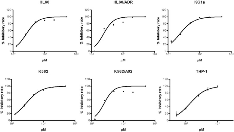
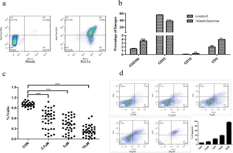
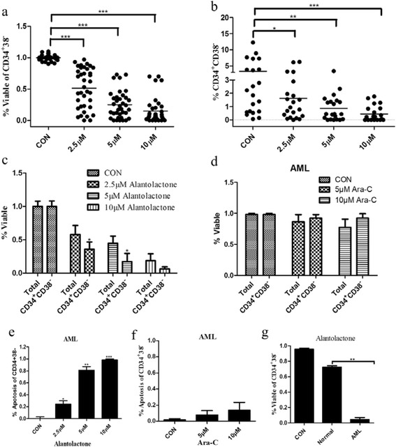
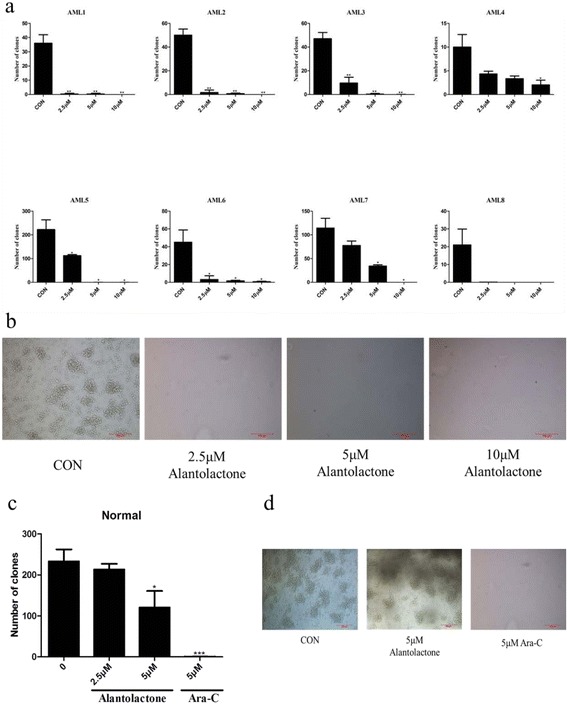
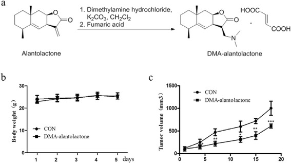
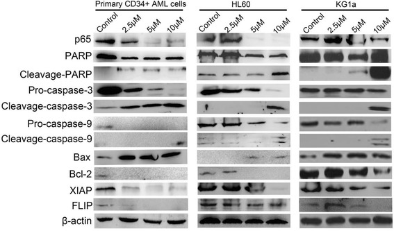
Results: The present study is the first to demonstrate that alantolactone, a prominent eudesmane-type sesquiterpene lactone, could specifically ablate LSCs from AML patient specimens. Furthermore, in comparison to the conventional chemotherapy drug, cytosine arabinoside (Ara-C), alantolactone showed superior effects of leukemia cytotoxicity while sparing normal hematopoietic cells. Alantolactone induced apoptosis with a dose-dependent manner by suppression of NF-kB and its downstream target proteins. DMA-alantolactone, a water-soluble prodrug of alantolactone, could suppress tumor growth in vivo.
Conclusions: Based on these results, we propose that alantolactone may represent a novel LSCs-targeted therapy and eudesmane-type sesquiterpene lactones offer a new scaffold for drug discovery towards anti-LSCs agents.
Keywords: Acute myeloid leukemia stem cells; Alantolactone; Apoptosis; KG1a.
Figures
Similar articles
-
The sesquiterpene lactone parthenolide induces apoptosis of human acute myelogenous leukemia stem and progenitor cells.Blood. 2005 Jun 1;105(11):4163-9. doi: 10.1182/blood-2004-10-4135. Epub 2005 Feb 1. Blood. 2005. PMID: 15687234 Free PMC article.
-
Antibody-Targeted Cyclodextrin-Based Nanoparticles for siRNA Delivery in the Treatment of Acute Myeloid Leukemia: Physicochemical Characteristics, in Vitro Mechanistic Studies, and ex Vivo Patient Derived Therapeutic Efficacy.Mol Pharm. 2017 Mar 6;14(3):940-952. doi: 10.1021/acs.molpharmaceut.6b01150. Epub 2017 Feb 14. Mol Pharm. 2017. PMID: 28146632
-
A New Strategy to Target Acute Myeloid Leukemia Stem and Progenitor Cells Using Chidamide, a Histone Deacetylase Inhibitor.Curr Cancer Drug Targets. 2015;15(6):493-503. doi: 10.2174/156800961506150805153230. Curr Cancer Drug Targets. 2015. PMID: 26282548
-
Characteristics of leukemic stem cells in acute leukemia and potential targeted therapies for their specific eradication.Cancer Drug Resist. 2022 May 5;5(2):344-367. doi: 10.20517/cdr.2021.140. eCollection 2022. Cancer Drug Resist. 2022. PMID: 35800375 Free PMC article. Review.
-
Therapeutic targeting of leukemia stem cells in acute myeloid leukemia.Front Oncol. 2023 Aug 3;13:1204895. doi: 10.3389/fonc.2023.1204895. eCollection 2023. Front Oncol. 2023. PMID: 37601659 Free PMC article. Review.
Cited by
-
KDM4 inhibitor SD49-7 attenuates leukemia stem cell via KDM4A/MDM2/p21CIP1 axis.Theranostics. 2022 Jun 21;12(11):4922-4934. doi: 10.7150/thno.71460. eCollection 2022. Theranostics. 2022. PMID: 35836814 Free PMC article.
-
Antineoplastic effects of CPPTL via the ROS/JNK pathway in acute myeloid leukemia.Oncotarget. 2017 Jun 13;8(24):38990-39000. doi: 10.18632/oncotarget.17166. Oncotarget. 2017. PMID: 28473664 Free PMC article.
-
Anticancer Activity of Curcumin-Loaded PLGA Nanoparticles on PC3 Prostate Cancer Cells.Iran J Pharm Res. 2017 Summer;16(3):868-879. Iran J Pharm Res. 2017. PMID: 29201078 Free PMC article.
-
microRNA-1246-containing extracellular vesicles from acute myeloid leukemia cells promote the survival of leukemia stem cells via the LRIG1-meditated STAT3 pathway.Aging (Albany NY). 2021 Apr 23;13(10):13644-13662. doi: 10.18632/aging.202893. Epub 2021 Apr 23. Aging (Albany NY). 2021. PMID: 33893245 Free PMC article.
-
Correction to: Alantolactone selectively ablates acute myeloid leukemia stem and progenitor cells.J Hematol Oncol. 2021 Apr 15;14(1):61. doi: 10.1186/s13045-021-01069-3. J Hematol Oncol. 2021. PMID: 33858468 Free PMC article. No abstract available.
References
LinkOut - more resources
Full Text Sources
Other Literature Sources
