. 2016 Oct:12:219-226.
doi: 10.1016/j.ebiom.2016.09.015. Epub 2016 Sep 17.

Bacterial Abscess Formation Is Controlled by the Stringent Stress Response and Can Be Targeted Therapeutically

Affiliations

Bacterial Abscess Formation Is Controlled by the Stringent Stress Response and Can Be Targeted Therapeutically

Sarah C Mansour et al. EBioMedicine. 2016 Oct.

Abstract

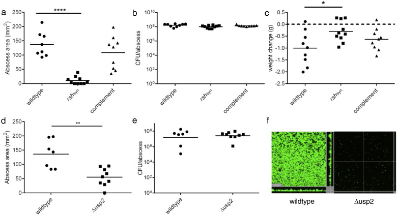
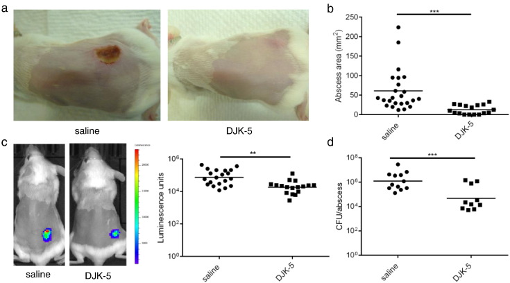
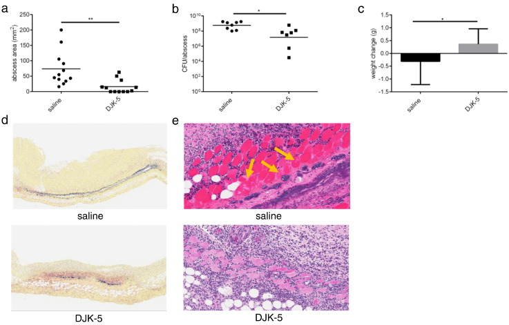
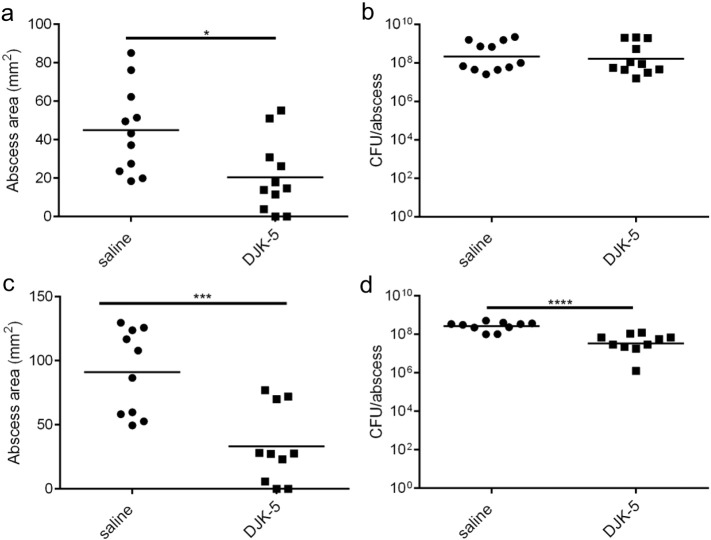
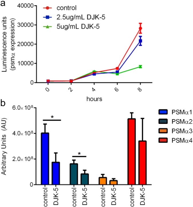
Cutaneous abscess infections are difficult to treat with current therapies and alternatives to conventional antibiotics are needed. Understanding the regulatory mechanisms that govern abscess pathology should reveal therapeutic interventions for these recalcitrant infections. Here we demonstrated that the stringent stress response employed by bacteria to cope and adapt to environmental stressors was essential for the formation of lesions, but not bacterial growth, in a methicillin resistant Staphylococcus aureus (MRSA) cutaneous abscess mouse model. To pharmacologically confirm the role of the stringent response in abscess formation, a cationic peptide that causes rapid degradation of the stringent response mediator, guanosine tetraphosphate (ppGpp), was employed. The therapeutic application of this peptide strongly inhibited lesion formation in mice infected with Gram-positive MRSA and Gram-negative Pseudomonas aeruginosa. Overall, we provide insights into the mechanisms governing abscess formation and a paradigm for treating multidrug resistant cutaneous abscesses.

Keywords: Cationic peptide; DJK-5; Pseudomonas aeruginosa; Staphylococcus aureus; ppGpp.


Figures

Fig. 1
Fig. 1
The stringent response was critical for S. aureus abscess lesion and biofilm formation. (a) Mice were infected subcutaneously with S. aureus HG001 wildtype, rshsyn and rshsyn complement strains. Lesions were measured 24 h post-infection using a caliper, n = 8–10/group. Statistical significance was determined using one-way ANOVA (****, p < 0.0001) (b) Bacteria were recovered 48 h post-infection from mouse abscesses infected with S. aureus HG001, ∆ rshsyn and complemented-rshsyn and then plated for enumeration, n = 9–10/group. (c) Weight loss/gain of infected mice was assessed by measuring the mouse weights pre-infection and 48 h post-infection, n = 9–10/group (*, p < 0.05). (d) Mice were infected subcutaneously with S. aureus Newman parent strain and its Δusp2 mutant. Lesion formation was measured 24 h post-infection using a caliper, n = 7–9/group. Statistical significance was determined using unpaired t-tests (**, p < 0.01). (e) Bacteria were recovered from Newman- and Δusp2- infected mice abscesses after 48 h and plated for enumeration, n = 7–9/group. (f) Newman and Δusp2 biofilms were grown in flow cells. After 72 h, bacteria were stained with Syto-9 (live bacteria stain) and propidium iodide (dead bacteria stain) prior to confocal imaging. The scale bar represents 30 μm in length and each image shows the xy, yz and xz dimensions. Two independent experiments were conducted for both animal studies and flow cell experiment.
Fig. 2
Fig. 2
Intraperitoneal application of DJK-5 impaired MRSA cutaneous lesion formation. (a) Mice were injected with 6 mg/kg of DJK-5 or saline as control via intraperitoneal injection prior to receiving subcutaneous injection with bioluminescent MRSA USA300. Representative images capturing dermonecrotic abscess lesions were taken 72 h post-infection. (b) Lesion sizes were measured three days post-infection using a caliper, n = 24 saline, n = 17 DJK-5. (c) Bioluminescent bacteria were imaged using In Vivo Imaging System (IVIS) and quantified using Living Image® Software, n = 20 saline, n = 17 DJK-5. (d) Five days post-infection, bacteria were recovered from saline or DJK-5 treated animals and plated for enumeration, n = 12 saline, n = 10 DJK-5. Three independent experiments were conducted and all comparisons were made using unpaired t tests; **, p < 0.01; ***, p < 0.001.
Fig. 3
Fig. 3
Intra-abscess application of DJK-5 impaired MRSA cutaneous injury. (a) Mice were injected with 3 mg/kg of DJK-5 or saline (control) via intra-abscess injection after subcutaneous infection with MRSA USA300. Abscess lesion sizes were measured using a caliper after 72 h, n = 12/group. (b) Bacterial enumeration from saline and DJK-5 intra-abscess-treated abscesses three days post-infection, n = 8 saline, n = 7 DJK-5. (c) Weight loss/gain of infected mice was assessed by measuring the mouse weights pre-infection and 72 h post-infection n = 12 saline, n = 11 DJK-5. (d) Representative Gram stain of saline and DJK-5 treated skin biopsies. (e) Hematoxylin and eosin stain of skin explants taken from saline and DJK-5 treated mice. Yellow arrows indicate suppurative myositis.
Fig. 4
Fig. 4
DJK-5 suppressed Gram-negative abscess lesion formation. (a) Mice were injected with 4 mg/kg of DJK-5 or saline (control) via intraperitoneal injection prior to subcutaneous infection with P. aeruginosa LESB58. Abscess lesion sizes were measured using a caliper after 72 h, n = 11/group. (b) Bacteria were recovered from saline and DJK-5 IP-treated animals and plated for enumeration three days post-infection, n = 11/group. (c) Mice were infected subcutaneously with P. aeruginosa LESB58 and then treated 2 h later with 4 mg/kg of DJK-5 via intra-abscess injection. Lesions were measured 72 h post-infection with a caliper, n = 10/group. (d) Bacteria were recovered from saline or DJK-5 intra-abscess treated animals and plated for enumeration, n = 10/group. All comparisons were made using unpaired t tests; *, p < 0.05; **, p < 0.01; ***, p < 0.001. All experiments were performed in triplicate.
Fig. 5
Fig. 5
DJK-5 targeted PSM production. (a) MRSA USA300 PSMα luminescence reporter strain was grown in the presence of 2.5 μg/mL, 5 μg/mL DJK-5 or water as a control. Luminescence activity was read using a VICTORX3 Multilabel Plate reader. (b) PSM production in culture filtrates of S. aureus MRSA USA300 treated with 1 μg/mL of DJK-5 or water as a control. All experiments were done in triplicate and all comparisons were done using unpaired t tests; *, p < 0.05.

References

    1. Aberg A., Shingler V., Balsalobre C. (p)ppGpp regulates type 1 fimbriation of Escherichia coli by modulating the expression of the site-specific recombinase FimB. Mol. Microbiol. 2006;60:1520–1533. - PubMed
    1. Attia A.S., Cassat J.E., Aranmolate S.O., Zimmerman L.J., Boyd K.L., Skaar E.P. Analysis of the Staphylococcus aureus abscess proteome identifies antimicrobial host proteins and bacterial stress responses at the host-pathogen interface. Pathog. Dis. 2013;69:36–48. - PMC - PubMed
    1. Berube B.J., Sampedro G.R., Otto M., Bubeck Wardenburg J. The psmα locus regulates production of Staphylococcus aureus alpha-toxin during infection. Infect. Immun. 2014;82:3350–3358. - PMC - PubMed
    1. Bolouri H., Savman K., Wang W., Thomas A., Maurer N., Dullaghan E., Fjell C.D., Ek C.J., Hagberg H., Hancock R.E.W. Innate defense regulator peptide 1018 protects against perinatal brain injury. Ann. Neurol. 2014;75:395–410. - PubMed
    1. Carpenter J.L. Klebsiella pulmonary infections: occurrence at one medical center and review. Rev. Infect. Dis. 1990;12:672–682. - PubMed

MeSH terms