

Bridging the gap between in vitro and in vivo RNA folding
- PMID: 27658939
- PMCID: PMC5269127
- DOI: 10.1017/S003358351600007X
Bridging the gap between in vitro and in vivo RNA folding
Abstract
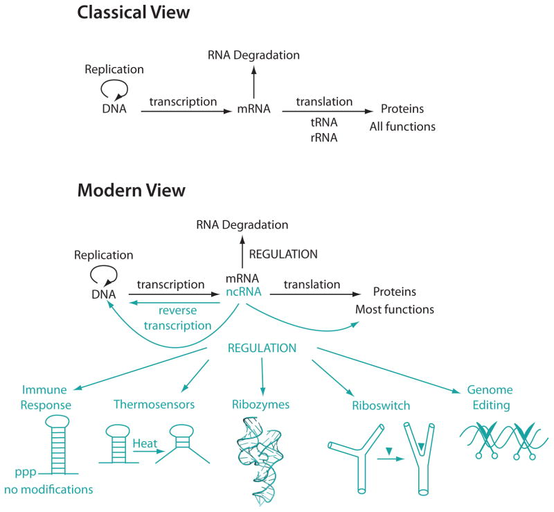
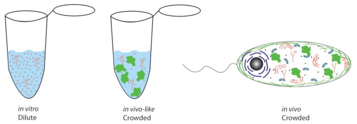
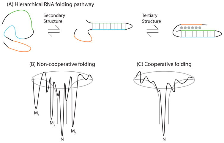
Deciphering the folding pathways and predicting the structures of complex three-dimensional biomolecules is central to elucidating biological function. RNA is single-stranded, which gives it the freedom to fold into complex secondary and tertiary structures. These structures endow RNA with the ability to perform complex chemistries and functions ranging from enzymatic activity to gene regulation. Given that RNA is involved in many essential cellular processes, it is critical to understand how it folds and functions in vivo. Within the last few years, methods have been developed to probe RNA structures in vivo and genome-wide. These studies reveal that RNA often adopts very different structures in vivo and in vitro, and provide profound insights into RNA biology. Nonetheless, both in vitro and in vivo approaches have limitations: studies in the complex and uncontrolled cellular environment make it difficult to obtain insight into RNA folding pathways and thermodynamics, and studies in vitro often lack direct cellular relevance, leaving a gap in our knowledge of RNA folding in vivo. This gap is being bridged by biophysical and mechanistic studies of RNA structure and function under conditions that mimic the cellular environment. To date, most artificial cytoplasms have used various polymers as molecular crowding agents and a series of small molecules as cosolutes. Studies under such in vivo-like conditions are yielding fresh insights, such as cooperative folding of functional RNAs and increased activity of ribozymes. These observations are accounted for in part by molecular crowding effects and interactions with other molecules. In this review, we report milestones in RNA folding in vitro and in vivo and discuss ongoing experimental and computational efforts to bridge the gap between these two conditions in order to understand how RNA folds in the cell.
Figures
References
-
- ALBERTS B, BRAY D, LEWIS J, ROBERTS K, WATSON JD. Molecular biology of the cell. 3. Garland Publishing; New York and London: 1994.
-
- ANDRONESCU M, CONDON A, TURNER DH, MATHEWS DH. The Determination of RNA Folding Nearest Neighbor Parameters. Methods in Molecular Biology. 2014;1097:45–70. - PubMed
-
- BAIRD NJ, WESTHOF E, QIN H, PAN T, SOSNICK TR. Structure of a folding intermediate reveals the interplay between core and peripheral elements in RNA folding. Journal of Molecular Biology. 2005;352:712–722. - PubMed
-
- BANERJEE AR, JAEGER JA, TURNER DH. Thermal unfolding of a group 1 ribozyme: the low-temperature transition is primarily disruption of the tertiary structure. Biochemistry. 1993;32(1):153–163. - PubMed
-
- BANERJEE AR, TURNER DH. The time dependence of chemical modification reveals slow steps in the folding of a Group I ribozyme. Biochemistry. 1995;34:6504–6512. - PubMed
Grants and funding
LinkOut - more resources
Full Text Sources
Other Literature Sources
Miscellaneous
