

When the Brain Takes 'BOLD' Steps: Real-Time fMRI Neurofeedback Can Further Enhance the Ability to Gradually Self-regulate Regional Brain Activation
- PMID: 27659118
- PMCID: PMC5953410
- DOI: 10.1016/j.neuroscience.2016.09.026
When the Brain Takes 'BOLD' Steps: Real-Time fMRI Neurofeedback Can Further Enhance the Ability to Gradually Self-regulate Regional Brain Activation
Abstract
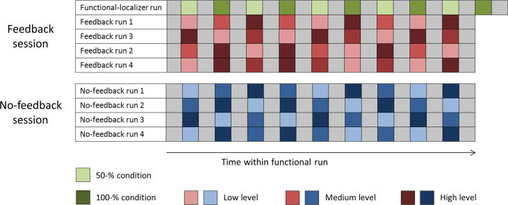
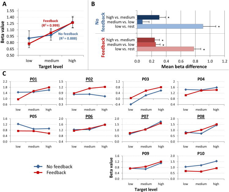
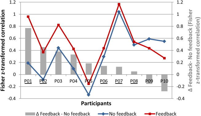
Brain-computer interfaces (BCIs) based on real-time functional magnetic resonance imaging (rtfMRI) are currently explored in the context of developing alternative (motor-independent) communication and control means for the severely disabled. In such BCI systems, the user encodes a particular intention (e.g., an answer to a question or an intended action) by evoking specific mental activity resulting in a distinct brain state that can be decoded from fMRI activation. One goal in this context is to increase the degrees of freedom in encoding different intentions, i.e., to allow the BCI user to choose from as many options as possible. Recently, the ability to voluntarily modulate spatial and/or temporal blood oxygenation level-dependent (BOLD)-signal features has been explored implementing different mental tasks and/or different encoding time intervals, respectively. Our two-session fMRI feasibility study systematically investigated for the first time the possibility of using magnitudinal BOLD-signal features for intention encoding. Particularly, in our novel paradigm, participants (n=10) were asked to alternately self-regulate their regional brain-activation level to 30%, 60% or 90% of their maximal capacity by applying a selected activation strategy (i.e., performing a mental task, e.g., inner speech) and modulation strategies (e.g., using different speech rates) suggested by the experimenters. In a second step, we tested the hypothesis that the additional availability of feedback information on the current BOLD-signal level within a region of interest improves the gradual-self regulation performance. Therefore, participants were provided with neurofeedback in one of the two fMRI sessions. Our results show that the majority of the participants were able to gradually self-regulate regional brain activation to at least two different target levels even in the absence of neurofeedback. When provided with continuous feedback on their current BOLD-signal level, most participants further enhanced their gradual self-regulation ability. Our findings were observed across a wide variety of mental tasks and across clinical MR field strengths (i.e., at 1.5T and 3T), indicating that these findings are robust and can be generalized across mental tasks and scanner types. The suggested novel parametric activation paradigm enriches the spectrum of current rtfMRI-neurofeedback and BCI methodology and has considerable potential for fundamental and clinical neuroscience applications.
Keywords: (gradual) self-regulation; (real-time) functional magnetic resonance imaging; brain-computer interface; cognitive strategies; mental tasks; neurofeedback.
Copyright © 2016 The Authors. Published by Elsevier Ltd.. All rights reserved.
Figures

Similar articles
-
Self-regulation of human brain activity using simultaneous real-time fMRI and EEG neurofeedback.Neuroimage. 2014 Jan 15;85 Pt 3:985-95. doi: 10.1016/j.neuroimage.2013.04.126. Epub 2013 May 11. Neuroimage. 2014. PMID: 23668969
-
Another kind of 'BOLD Response': answering multiple-choice questions via online decoded single-trial brain signals.Prog Brain Res. 2009;177:275-92. doi: 10.1016/S0079-6123(09)17719-1. Prog Brain Res. 2009. PMID: 19818908
-
Real-time fMRI and its application to neurofeedback.Neuroimage. 2012 Aug 15;62(2):682-92. doi: 10.1016/j.neuroimage.2011.10.009. Epub 2011 Oct 14. Neuroimage. 2012. PMID: 22019880 Review.
-
Self-regulation of the anterior insula: Reinforcement learning using real-time fMRI neurofeedback.Neuroimage. 2014 Mar;88:113-24. doi: 10.1016/j.neuroimage.2013.10.069. Epub 2013 Nov 11. Neuroimage. 2014. PMID: 24231399
-
Real-time fMRI brain computer interfaces: self-regulation of single brain regions to networks.Biol Psychol. 2014 Jan;95:4-20. doi: 10.1016/j.biopsycho.2013.04.010. Epub 2013 May 1. Biol Psychol. 2014. PMID: 23643926 Review.
Cited by
-
Success and failure of controlling the real-time functional magnetic resonance imaging neurofeedback signal are reflected in the striatum.Brain Behav. 2019 Mar;9(3):e01240. doi: 10.1002/brb3.1240. Epub 2019 Feb 20. Brain Behav. 2019. PMID: 30790474 Free PMC article.
-
Quality and denoising in real-time functional magnetic resonance imaging neurofeedback: A methods review.Hum Brain Mapp. 2020 Aug 15;41(12):3439-3467. doi: 10.1002/hbm.25010. Epub 2020 Apr 25. Hum Brain Mapp. 2020. PMID: 32333624 Free PMC article. Review.
-
Consensus on the reporting and experimental design of clinical and cognitive-behavioural neurofeedback studies (CRED-nf checklist).Brain. 2020 Jun 1;143(6):1674-1685. doi: 10.1093/brain/awaa009. Brain. 2020. PMID: 32176800 Free PMC article.
-
Reinforcement and Punishment Shape the Learning Dynamics in fMRI Neurofeedback.Front Hum Neurosci. 2020 Jul 24;14:304. doi: 10.3389/fnhum.2020.00304. eCollection 2020. Front Hum Neurosci. 2020. PMID: 32792929 Free PMC article.
-
A feasibility study of goal-directed network-based real-time fMRI neurofeedback for anhedonic depression.Front Psychiatry. 2023 Dec 5;14:1253727. doi: 10.3389/fpsyt.2023.1253727. eCollection 2023. Front Psychiatry. 2023. PMID: 38125285 Free PMC article.
References
-
- Birbaumer N., Ghanayim N., Hinterberger T., Iversen I., Kotchoubey B., Kübler A., Perelmouter J., Taub E., Flor H. A spelling device for the paralysed. Nature. 1999;398:297–298. - PubMed
Publication types
MeSH terms
Substances
Grants and funding
LinkOut - more resources
Full Text Sources
Other Literature Sources
Medical
