

Cleavage of E-Cadherin Contributes to Defective Barrier Function in Neosquamous Epithelium
- PMID: 27659669
- PMCID: PMC5290423
- DOI: 10.1007/s10620-016-4315-y
Cleavage of E-Cadherin Contributes to Defective Barrier Function in Neosquamous Epithelium
Abstract
Background: After ablation of Barrett's esophagus (BE), the esophagus heals with neosquamous epithelium (NSE). Despite normal endoscopic appearance, NSE exhibits defective barrier function with similarities to defects noted in the distal esophageal epithelium in patients with gastroesophageal reflux disease (GERD).
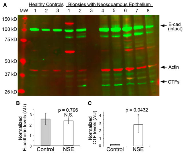
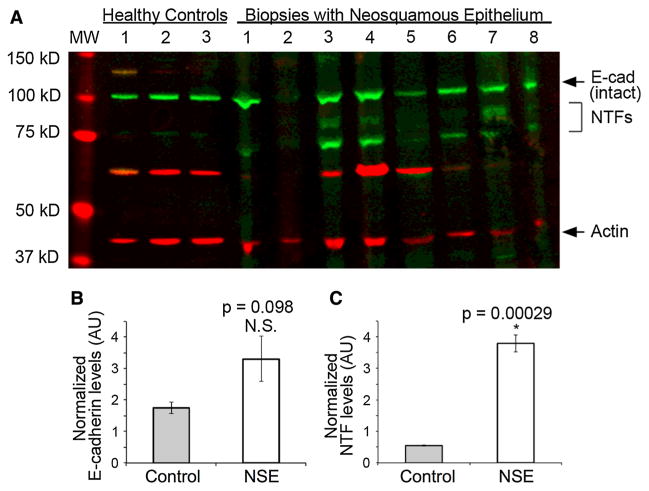
Aim: To determine whether patients with NSE, unlike patients with healthy esophageal epithelium, have C-terminal fragments (CTFs) of e-cad detectable on tissue biopsy. Secondly, to determine whether patients with NSE have elevated levels of N-terminal fragments (NTFs) of e-cad in the serum.
Methods: Fifteen patients with ablated long-segment BE, who had healing with formation of NSE, were enrolled in this pilot study. Western blots for CTFs and NTFs were performed on biopsies of NSE. Venous blood was obtained to assess levels of NTFs. Endoscopic distal esophageal biopsies from patients without esophageal disease served as tissue controls. Control blood samples were obtained from healthy subjects.
Results: Blots of NSE were successful in 14/15 patients, and all 14 (100 %) had a 35-kD CTF of e-cad, while CTFs were absent in healthy control tissues. Despite CTFs in NSE, serum NTFs of e-cad in NSE were similar to controls, p > 0.05. However, unlike healthy controls, blots of NSE also showed NTFs with molecular weights of 70-90 kD.
Conclusions: Cleavage of e-cad, as evidenced by the presence of CTFs and NTFs on biopsy, contributes to defective barrier function in NSE. However, unlike findings reported in GERD patients, serum NTFs are not elevated in NSE patients. This difference may reflect poor absorption with tissue entrapment of NTFs in previously ablated areas with poorly perfused NSE.
Keywords: Barrett’s esophagus; ELISA; Esophageal permeability; Radiofrequency ablation; Western blot.
Conflict of interest statement
Dr. Runge has no conflicts to declare. RC Orlando and Z Djukic have a patent for using the identification of fragments of e-cadherin for the diagnosis of GERD.
Figures
Similar articles
-
Fragments of e-Cadherin as Biomarkers of Non-erosive Reflux Disease.Dig Dis Sci. 2018 Mar;63(3):628-635. doi: 10.1007/s10620-017-4815-4. Epub 2017 Oct 25. Dig Dis Sci. 2018. PMID: 29071486 Free PMC article.
-
MicroRNA profile in neosquamous esophageal mucosa following ablation of Barrett's esophagus.World J Gastroenterol. 2017 Aug 14;23(30):5508-5518. doi: 10.3748/wjg.v23.i30.5508. World J Gastroenterol. 2017. PMID: 28852310 Free PMC article.
-
Defective barrier function in neosquamous epithelium.Am J Gastroenterol. 2013 Mar;108(3):386-91. doi: 10.1038/ajg.2012.440. Epub 2013 Jan 15. Am J Gastroenterol. 2013. PMID: 23318477 Free PMC article.
-
Endoscopic therapy using radiofrequency ablation for esophageal dysplasia and carcinoma in Barrett's esophagus.Gastrointest Endosc Clin N Am. 2010 Jan;20(1):55-74, vi. doi: 10.1016/j.giec.2009.07.007. Gastrointest Endosc Clin N Am. 2010. PMID: 19951794 Review.
-
Diagnosis and Management of Low-Grade Dysplasia in Barrett's Esophagus: Expert Review From the Clinical Practice Updates Committee of the American Gastroenterological Association.Gastroenterology. 2016 Nov;151(5):822-835. doi: 10.1053/j.gastro.2016.09.040. Epub 2016 Oct 1. Gastroenterology. 2016. PMID: 27702561 Review.
Cited by
-
Does radiofrequency ablation of the lower oesophagus allow for clonal expansion of highly mutated neosquamous epithelium?BMJ Oncol. 2023 Oct 29;2(1):e000089. doi: 10.1136/bmjonc-2023-000089. eCollection 2023. BMJ Oncol. 2023. PMID: 39886511 Free PMC article.
-
Novel histologic score predicts recurrent intestinal metaplasia after successful endoscopic eradication therapy.Dis Esophagus. 2023 Apr 29;36(5):doac078. doi: 10.1093/dote/doac078. Dis Esophagus. 2023. PMID: 36446594 Free PMC article.
-
Is post-ablation neo-squamous epithelium genomically predisposed to malignant progression?BMJ Oncol. 2023 Oct 29;2(1):e000183. doi: 10.1136/bmjonc-2023-000183. eCollection 2023. BMJ Oncol. 2023. PMID: 39886506 Free PMC article. No abstract available.
-
Fragments of e-Cadherin as Biomarkers of Non-erosive Reflux Disease.Dig Dis Sci. 2018 Mar;63(3):628-635. doi: 10.1007/s10620-017-4815-4. Epub 2017 Oct 25. Dig Dis Sci. 2018. PMID: 29071486 Free PMC article.
-
Electric Field Based Dressing Disrupts Mixed-Species Bacterial Biofilm Infection and Restores Functional Wound Healing.Ann Surg. 2019 Apr;269(4):756-766. doi: 10.1097/SLA.0000000000002504. Ann Surg. 2019. PMID: 29099398 Free PMC article.
References
-
- Shaheen NJ, Richter JE. Barrett’s oesophagus. Lancet. 2009;373:850–861. - PubMed
-
- Spechler SJ. Barrett’s esophagus and esophageal adenocarcinoma: pathogenesis, diagnosis, and therapy. Med Clin N Am. 2002;86:1423–1445. - PubMed
-
- Spechler SJ. Barrett’s esophagus. In: Orlando RC, editor. Gastroesophageal Reflux Disease. New York, NY: CRC Press; 2000. pp. 219–258.
-
- Gerson LB, Shetler K, Triadafilopoulos G. Prevalence of Barrett’s esophagus in asymptomatic individuals. Gastroenterology. 2002;123:461–467. - PubMed
-
- Rex DK, Cummings OW, Shaw M, et al. Screening for Barrett’s esophagus in colonoscopy patients with and without heartburn. Gastroenterology. 2003;125:1670–1677. - PubMed
Publication types
MeSH terms
Substances
Grants and funding
LinkOut - more resources
Full Text Sources
Other Literature Sources
Miscellaneous
