Review
. 2016 Oct;8(10):1399-1413.
doi: 10.2217/epi-2016-0056. Epub 2016 Sep 23.

Establishment and functions of DNA methylation in the germline

Affiliations
Review

Establishment and functions of DNA methylation in the germline

Kathleen R Stewart et al. Epigenomics. 2016 Oct.

Abstract

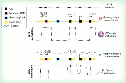
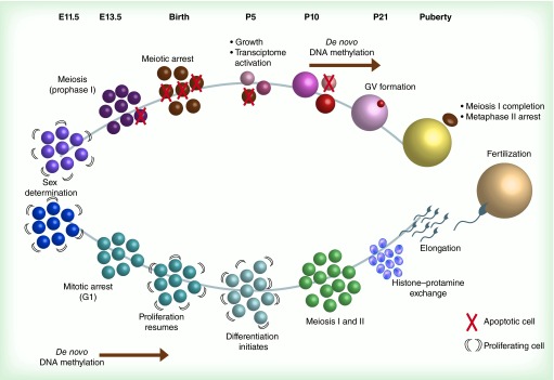
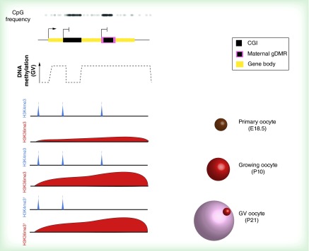
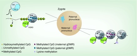
Epigenetic modifications established during gametogenesis regulate transcription and other nuclear processes in gametes, but also have influences in the zygote, embryo and postnatal life. This is best understood for DNA methylation which, established at discrete regions of the oocyte and sperm genomes, governs genomic imprinting. In this review, we describe how imprinting has informed our understanding of de novo DNA methylation mechanisms, highlight how recent genome-wide profiling studies have provided unprecedented insights into establishment of the sperm and oocyte methylomes and consider the fate and function of gametic methylation and other epigenetic modifications after fertilization.

Keywords: DNA methylation; embryo; imprinting; oogenesis; spermatogenesis; transgenerational inheritance.


Conflict of interest statement

Financial & competing interests disclosure KR Stewart was supported by a studentship from the Cambridge Overseas Trusts. L Veselovska was supported by a Babraham Institute: Cambridge University studentship. Work in G Kelsey's laboratory is funded by the Biotechnology and Biological Sciences Research Council and the Medical Research Council. The authors have no other relevant affiliations or financial involvement with any organization or entity with a financial interest in or financial conflict with the subject matter or materials discussed in the manuscript apart from those disclosed. No writing assistance was utilized in the production of this manuscript.

Figures

<b>Figure 1.</b>
Figure 1.. The oocyte and sperm methylomes.
Methylated regions in the GV oocyte (top) tightly correspond with active transcription units in growing oocytes, initiating from both CGI and non-CGI transcription start sites. CGIs that become de novo methylated in oocytes, including maternally imprinted gDMRs, tend to be within active transcription units and CpG-rich. In the male germline, regions that were both transcribed and untranscribed in prospermatogonia are methylated in mature sperm (bottom). Promoters of genes with weak or no expression in prospermatogonia display intermediate methylation in sperm, from either heterogeneous methylation of these regions in the germ cell pool or methylation heterogeneity in individual CpGs at these genomic regions. Paternally imprinted gDMRs tend to be less CpG-dense than maternally imprinted gDMRs. Notably, in either germline, transcription levels past a low threshold result in full methylation of the transcription unit, but active promoters at CGIs are protected from DNA methylation. Arrows, active transcription start sites; crosses, inactive transcription start sites. CGI: CpG island; gDMR: Germline differentially methylated region; GV: Germinal vesicle.
<b>Figure 2.</b>
Figure 2.. Nuclear and epigenetic dynamics in gametogenesis: temporal and developmental perspectives.
In mice, sex determination occurs in the gonadal ridge between E10.5 and E12.5. At E13.5, primordial germ cells (PGCs) in both male and female developing gonads stop proliferating: female PGCs enter meiosis, while male PGCs (called prospermatogonia or gonocytes) arrest in mitotic G1 and undergo de novo DNA methylation between E13.5 and birth. By birth (approximately 19 days after conception), female PGCs have become primary oocytes, and arrest at the diplotene stage of meiotic prophase I. Many oocytes undergo apoptosis at this time, and oocytes continue to die throughout oogenesis. Male gametes, now spermatogonia, resume proliferation. At P5, oocytes begin expanding their cytoplasmic volume, and oocyte-specific transcription units also become active. This oocyte growth is asynchronous, with some oocytes growing faster than others. Spermatogonia begin to differentiate. De novo methylation begins in oocytes at around P10, or when an oocyte reaches at least 40–45 μm in diameter. Spermatogonia undergo meiosis at this time. Histone-protamine exchange occurs in round spermatids at P21, which later elongate into mature spermatids. At P21, the oocyte methylome is established, and GVs form. Upon ovulation, oocytes become developmentally competent by completing meiosis I and arresting in metaphase II, while extruding the first polar body. GV: Germinal vesicle.
<b>Figure 3.</b>
Figure 3.. Chromatin modulation accompanies establishment of the oocyte DNA methylome.
CGIs and maternally imprinted gDMRs that gain methylation in the oocyte tend to be within active transcription units, but not all transcribed CGIs acquire DNA methylation (top). In the scheme shown, the yellow bar at the top represents a single transcription unit that contains two intragenic CGIs that are initially both marked with H3K4me3 in primary oocytes at E18.5 (these CGIs may be active promoters in other cell types). In growing (P10) oocytes, most CGIs destined for DNA methylation retain this H3K4me3 and, compared with CGIs that resist DNA methylation, they also become enriched in H3K36me3. Presumed loss of H3K4me3 sometime between P10 and P21 then permits DNA methylation establishment at these loci by the GV oocyte stage. Hypothesized profiles. CGI: CpG island; gDMR: Germline differentially methylated region; GV: Germinal vesicle.
<b>Figure 4.</b>
Figure 4.. Gametic epigenomes in the zygote.
Following fertilization, most of the paternal genome is actively demethylated, during which time it is characterized by widespread hydroxymethylation. The few canonical nucleosomes present in mature sperm are predicted to persist in the paternal pronucleus, but in the majority of the genome protamines (green circles) are exchanged for maternally provided histones containing the H3 variant H3.3. The maternal genome is largely not subject to either of these remodeling events. Nucleosomes marked with H3K9me2 recruit PGC7/STELLA, which protects the maternal genome from demethylation. The same mechanism protects DNA methylation over at least two paternally imprinted gDMRs. gDMR: Germline differentially methylated region; PGC: Primordial germ cell.

References

    1. Jones PA. Functions of DNA methylation: islands, start sites, gene bodies and beyond. Nat. Rev. Genet. 2012;13(7):484–492. - PubMed
    1. Chen T, Li E. Structure and function of eukaryotic DNA methyltransferases. Curr. Top. Dev. Biol. 2004;60:55–89. - PubMed
    1. Hitt MM, Wu TL, Cohen G, Linn S. De novo and maintenance DNA methylation by a mouse plasmacytoma cell DNA methyltransferase. J. Biol. Chem. 1988;263(9):4392–4399. - PubMed
    1. Pradhan S, Bacolla A, Wells RD, Roberts RJ. Recombinant human DNA (cytosine-5) methyltransferase: I. Expression, purification, and comparison of de novo and maintenance methylation. J. Biol. Chem. 1999;274(46):33002–33010. - PubMed
    1. Arita K, Ariyoshi M, Tochio H, Nakamura Y, Shirakawa M. Recognition of hemi-methylated DNA by the SRA protein UHRF1 by a base-flipping mechanism. Nature. 2008;455(7214):818–821. - PubMed

Publication types