Review
. 2016 Oct;32(10):1823-32.
doi: 10.1007/s00381-016-3125-z. Epub 2016 Sep 20.

PET and SPECT studies in children with hemispheric low-grade gliomas

Affiliations
Review

PET and SPECT studies in children with hemispheric low-grade gliomas

Csaba Juhász et al. Childs Nerv Syst. 2016 Oct.

Abstract

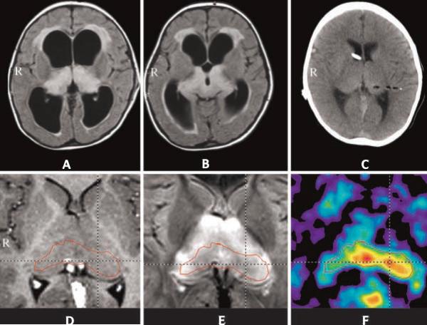
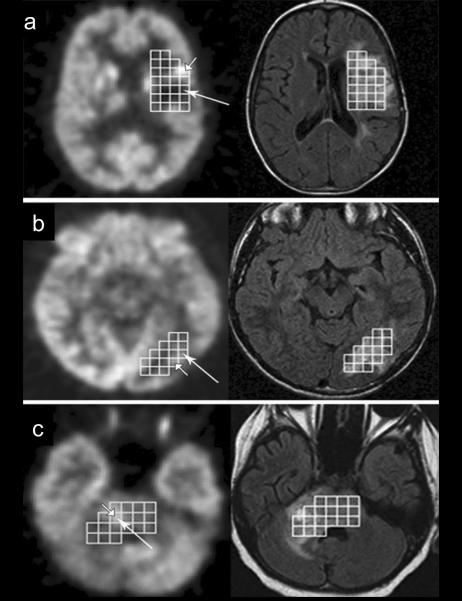
Molecular imaging is playing an increasing role in the pretreatment evaluation of low-grade gliomas. While glucose positron emission tomography (PET) can be helpful to differentiate low-grade from high-grade tumors, PET imaging with amino acid radiotracers has several advantages, such as better differentiation between tumors and non-tumorous lesions, optimized biopsy targeting, and improved detection of tumor recurrence. This review provides a brief overview of single-photon emission computed tomography (SPECT) studies followed by a more detailed review of the clinical applications of glucose and amino acid PET imaging in low-grade hemispheric gliomas. We discuss key differences in the performance of the most commonly utilized PET radiotracers and highlight the advantage of PET/MRI fusion to obtain optimal information about tumor extent, heterogeneity, and metabolism. Recent data also suggest that simultaneous acquisition of PET/MR images and the combination of advanced MRI techniques with quantitative PET can further improve the pretreatment and post-treatment evaluation of pediatric brain tumors.

Keywords: Amino acid; Glucose; Low-grade; MRI; Pediatric glioma; Positron emission tomography.


Figures

Figure 1
Figure 1
Comparison of fluid-attenuated inversion recovery (FLAIR) MR (A) and FDOPA-PET/MR fusion images (B) in a patient with a WHO grade 2 diffuse astrocytoma. FDOPA did not show any tracer uptake in this low-grade glioma (from Morana et al. [27]).
Figure 2
Figure 2
MR and O-(2-[18F]fluoroethyl)-L-tyrosine (FET)-PET images of a 2-year old girl with bithalamic lesions, which appeared as hyperintense signals on FLAIR images (A, B). Postoperative CT scan (C) demonstrated the biopsy tract contrasting with air. MRI after shunting showed the thalamic region on the post-contrast T1-weighted image without pathologic enhancement (D) and symmetrical hyperintensity on the FLAIR sequence (E). The corresponding FET-PET demonstrated increased uptake (tumor/cortex ratio 1.6) in the dorsal midline of the bithalamic region as well as the left pulvinar. Histopathology indicated low-grade astrocytoma (from Messinger-Jünger et al. [24]).
Figure 3
Figure 3
Voxel-wise comparison of FDG-PET (left side) and FLAIR images (right side) with MR spectroscopic imaging voxels in 3 patients (a, b, c) with gliomas. Voxels with maximum choline/N-acetyl-aspartate (Cho/NAA) ratio are indicated by long arrows, while the brightest voxel with low Cho/NAA is shown by short arrow. a: No agreement between FDG-PET and MRS voxel selection. b and c: agreement between FDG-PET and MRS images. b: Selected voxels are adjacent to one another and c: the same voxel was chosen by both FDG-PET and MRS (from Hipp et al. [12]).
Figure 4
Figure 4
Stereotactic comparison of MET-PET (left) and contrast-enhanced MRI (right) of a metabolically heterogeneous, non-enhancing glioma. Based on MRI, biopsy was preferred to resection because of the deep tumor infiltration. MET-PET showed heterogeneous tracer uptake with a focus of maximum metabolism used as a biopsy target (arrow 1); the tissue obtained from this area showed anaplastic features, while biopsy from the area with low tracer uptake (and hypointensity on MRI, arrow 2) yielded non-tumoral tissue (modified from Pirotte et al. [35]).
Figure 5
Figure 5
Comparison of FLAIR (A), post-contrast T1 (B), perfusion-weighted imaging (PWI) (C) and α-[11C]methyl-L-tryptophan (AMT)-PET images (D) of a low-grade glioma. FLAIR image showed a hyperintense mass in the right pre-central region, with no contrast enhancement and normal blood volume on PWI. AMT-PET showed high tryptophan uptake in the tumor mass, a common finding in low-grade gliomas.
Figure 6
Figure 6
Postoperative MRI (a) and 11C-methionine (MET)-PET (b) of an ependymoma. MRI was performed 24 hours after surgery and MET-PET was done 5 days after resection. On MR images two linear signals were seen along the resection cavity (arrow), suggesting a questionable residual tumor. MET-PET showed high tracer uptake confirming a residual tumor mass (from Pirotte et al. [34]).

Similar articles

Cited by

References

    1. Alkonyi B, Mittal S, Zitron I, et al. Increased tryptophan transport in epileptogenic dysembryoplastic neuroepithelial tumors. J Neurooncol. 2012;107:365–72. - PMC - PubMed
    1. Bagni B, Pinna L, Tamarozzi R, et al. SPET imaging of intracranial tumours with 99Tcm-sestamibi. Nucl Med Commun. 1995;16:258–64. - PubMed
    1. Batista CE, Juhasz C, Muzik O, et al. Imaging correlates of differential expression of indoleamine 2,3-dioxygenase in human brain tumors. Mol Imaging Biol. 2009;11:460–6. - PMC - PubMed
    1. Black KL, Hawkins RA, Kim KT, Becker DP, Lerner C, Marciano D. Use of thallium-201 SPECT to quantitate malignancy grade of gliomas. J Neurosurg. 1989;71:342–6. - PubMed
    1. Borgwardt L, Larsen HJ, Pedersen K, Højgaard L. Practical use and implementation of PET in children in a hospital PET centre. Eur J Nucl Med Mol Imaging. 2003;30:1389–1397. - PubMed

MeSH terms

Substances

LinkOut - more resources