. 2017 Jun;30(6):878-885.
doi: 10.5713/ajas.16.0515. Epub 2016 Sep 19.

Mammary alveolar cell as in vitro evaluation system for casein gene expression involved in glucose level

Affiliations

Mammary alveolar cell as in vitro evaluation system for casein gene expression involved in glucose level

Young Tae Heo et al. Asian-Australas J Anim Sci. 2017 Jun.

Abstract

Objective: Glucose is an essential fuel in the energy metabolism and synthesis pathways of all mammalian cells. In lactating animals, glucose is the major precursor for lactose and is a substrate for the synthesis of milk proteins and fat in mammary secretory (alveolar) epithelial cells. However, clear utilization of glucose in mammary cells during lactogenesis is still unknown, due to the lack of in vitro analyzing models. Therefore, the objective of this study was to test the reliability of the mammary alveolar (MAC-T) cell as an in vitro study model for glucose metabolism and lactating system.

Methods: Undifferentiated MAC-T cells were cultured in three types of Dulbecco's modified Eagle's medium with varying levels of glucose (no-glucose: 0 g/L, low-glucose: 1 g/L, and high-glucose: 4.5 g/L) for 8 d, after which differentiation to casein secretion was induced. Cell proliferation and expression levels of apoptotic genes, Insulin like growth factor-1 (IGF1) receptor, oxytocin receptor, αS1, αS2, and β casein genes were analyzed at 1, 2, 4, and 8 d after differentiation.

Results: The proliferation of MAC-T cells with high-glucose treatment was seen to be significantly higher. Expression of apoptotic genes was not affected in any group. However, expression levels of the mammary development related gene (IGF1 receptor) and lactation related gene (oxytocin receptor) were significantly higher in the low-glucose group. Expressions of αS1-casein, αS2-casein, and β-casein were also higher in the low-glucose treated group as compared to that in the no-glucose and high-glucose groups.

Conclusion: The results demonstrated that although a high-glucose environment increases cell proliferation in MAC-T cells, a low-glucose treatment to MAC-T cells induces higher expression of casein genes. Our results suggest that the MAC-T cells may be used as an in vitro model to analyze mammary cell development and lactation connected with precise biological effects.

Keywords: Casein; Glucose; Insulin-like Growth Factor-1 (IGF-1); Mammary alveolar (MAC-T) Cell; Oxytocin.


Conflict of interest statement

CONFLICT OF INTEREST

We certify that there is no conflict of interest with any financial organization regarding the material discussed in the manuscript.

Figures

Figure 1
Figure 1
Effects of glucose level on the proliferation and apoptosis of MAC-T cells. The cells were cultures in none, low and high (0, 1, and 4.5 g/L) glucose DMEM media. MAC-T cells which treated different level of glucose were observed by microscope on 1, 2, 4, and 8 d (A). The cell proliferations of the treatment groups before (B) and after (C) differentiation induction was analyzed by cell counting. Apoptotic effects by different glucose level were determined by real-time, reverse transcription-PCR and GAPDH was use as the endogenous housekeeping gene (D, E). Expression of the apoptosis-related genes caspase8 (D) Bcl-xL and Bax (E) in MAC-T cells was measured at 1, 2, 4, and 8 d after induction of differentiation. Results are expressed as the Bcl-xL:Bax mRNA ratios (E). Each experiment was repeated 3 times. All values are expressed as mean± standard error of the mean. MAC-T, mammary alveolar; DMEM, Dulbecco’s modified Eagle’s medium; PCR, polymerase chain reaction; GAPDH, glyceraldehyde 3-phosphate dehydrogenase. Each experimental group indicated a–c to significant differences between each treatment for same days (p<0.05).
Figure 2
Figure 2
Effects of glucose level on the expression of glucose transporters in differentiated MAC-T cells. Expression levels of mammary gland specific glucose transporters (1, 4, 8, and 12) genes were determined by real-time, reverse transcription-PCR and GAPDH was used as the endogenous housekeeping gene. Expressions of GLUT 1 (A), 4 (B), 8 (C), and 12 (D) genes in MAC-T cells was measured at 1, 2, 4 and 8 d after induction of differentiation. Each experiment was repeated 3 times. All values are expressed as mean±standard error of the mean. MAC-T, mammary alveolar; PCR, polymerase chain reaction; GAPDH, glyceraldehyde 3-phosphate dehydrogenase; GLUT, glucose transporter.
Figure 3
Figure 3
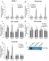
Effects of glucose level on the expression of bovine IGF-1 receptor, oxytocin receptor, αS1-casein, αS2-casein, and β-casein genes in differentiated MAC-T cells. Expression levels of genes were determined by real-time, reverse transcription-PCR and GAPDH was used as the endogenous housekeeping gene. Expression of the mammary cell development related gene IGF-1 receptor (A), and lactation related gene Oxytocin receptor (B) in MAC-T cells was measured at 1, 2, 4, and 8 d after induction of differentiation. Expressions of bovine αS1-casein (C), αS2-casein (D), and β-casein (E) genes were analyzed at 1, 2, 4, and 8 d after induction of differentiation. Expression of bovine total casein in cultured media (F) was identified at 4 d after induction of differentiation. Each experiment was repeated 3 times. All values are expressed as mean±standard error of the mean. IGF-1, insulin like growth factor-1; MAC-T, mammary alveolar; PCR, polymerase chain reaction; GAPDH, glyceraldehyde 3-phosphate dehydrogenase. Each experimental group indicated a–c to significant differences between each treatment for same days (p<0.05).

References

    1. Cardenas H, Carvajal A, Utreras E, et al. Lactation inhibits the potentiating effect of galanin upon the GnRH-induced LH release observed in diestrous-1 rat. Biol Res. 1998;31:351–8. - PubMed
    1. Threadgold LC, Kuhn NJ. Glucose-6-phosphate hydrolysis by lactating rat mammary gland. Int J Biochem. 1979;10:683–5. - PubMed
    1. Kuhn NJ, Carrick DT, Wilde CJ. Lactose synthesis: the possibilities of regulation. J Dairy Sci. 1980;63:328–36. - PubMed
    1. Stacey A, Schnieke A, Kerr M, et al. Lactation is disrupted by alpha-lactalbumin deficiency and can be restored by human alpha-lactalbumin gene replacement in mice. Proc Natl Acad Sci USA. 1995;92:2835–9. - PMC - PubMed
    1. Xiao CT, Cant JP. Relationship between glucose transport and metabolism in isolated bovine mammary epithelial cells. J Dairy Sci. 2005;88:2794–805. - PubMed

LinkOut - more resources