

68Ga-PSMA-11 PET Imaging of Response to Androgen Receptor Inhibition: First Human Experience
- PMID: 27660139
- PMCID: PMC5209643
- DOI: 10.2967/jnumed.116.181800
68Ga-PSMA-11 PET Imaging of Response to Androgen Receptor Inhibition: First Human Experience
Abstract
The purpose of this work was to evaluate the effect of androgen receptor (AR) inhibition on prostate-specific membrane antigen (PSMA) uptake imaged using 68Ga-PSMA-11 PET in a mouse xenograft model and in a patient with castration-sensitive prostate cancer.
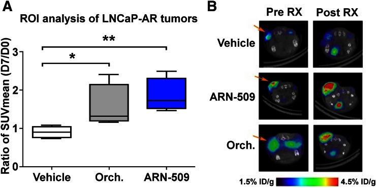
Methods: We imaged 3 groups of 4 mice bearing LNCaP-AR xenografts before and 7 d after treatment with ARN-509, orchiectomy, or control vehicle. Additionally, we imaged one patient with castration-sensitive prostate cancer before and 4 wk after treatment with androgen deprivation therapy (ADT). Uptake on pre- and posttreatment imaging was measured and compared.
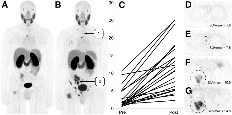
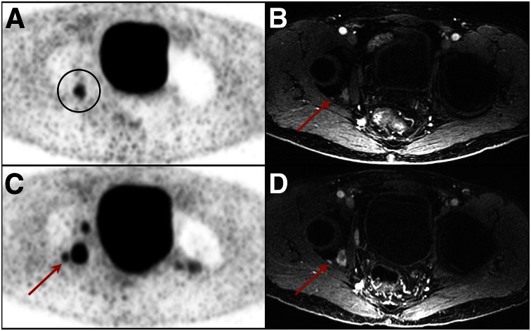
Results: PSMA uptake increased 1.5- to 2.0-fold in the xenograft mouse model after treatment with both orchiectomy and ARN-509 but not with vehicle. Patient imaging demonstrated a 7-fold increase in PSMA uptake after the initiation of ADT. Thirteen of 22 lesions in the imaged patient were visualized on PSMA PET only after treatment with ADT.
Conclusion: Inhibition of the AR can increase PSMA expression in prostate cancer metastases and increase the number of lesions visualized using PSMA PET. The effect seen in cell and animal models can be recapitulated in humans. A better understanding of the temporal changes in PSMA expression is needed to leverage this effect for both improved diagnosis and improved therapy.
Keywords: PET; PSMA PET; androgen receptor; oncology: GU; prostate cancer.
© 2017 by the Society of Nuclear Medicine and Molecular Imaging.
Figures
References
-
- Israeli RS, Powell CT, Corr JG, Fair WR, Heston WD. Expression of the prostate-specific membrane antigen. Cancer Res. 1994;54:1807–1811. - PubMed
-
- Silver DA, Pellicer I, Fair WR, Heston WD, Cordon-Cardo C. Prostate-specific membrane antigen expression in normal and malignant human tissues. Clin Cancer Res. 1997;3:81–85. - PubMed
-
- Hijazi S, Meller B, Leitsmann C, et al. Pelvic lymph node dissection for nodal oligometastatic prostate cancer detected by 68Ga-PSMA-positron emission tomography/computerized tomography. Prostate. 2015;75:1934–1940. - PubMed
-
- Ceci F, Uprimny C, Nilica B, et al. 68Ga-PSMA PET/CT for restaging recurrent prostate cancer: which factors are associated with PET/CT detection rate? Eur J Nucl Med Mol Imaging. 2015;42:1284–1294. - PubMed
Publication types
MeSH terms
Substances
Grants and funding
LinkOut - more resources
Full Text Sources
Other Literature Sources
Research Materials
Miscellaneous