

Green synthesis of silver nanoparticles using Pimpinella anisum seeds: antimicrobial activity and cytotoxicity on human neonatal skin stromal cells and colon cancer cells
- PMID: 27660438
- PMCID: PMC5019319
- DOI: 10.2147/IJN.S113193
Green synthesis of silver nanoparticles using Pimpinella anisum seeds: antimicrobial activity and cytotoxicity on human neonatal skin stromal cells and colon cancer cells
Abstract
Background: The present study focused on a simple and eco-friendly method for the synthesis of silver nanoparticles (AgNPs) with multipurpose anticancer and antimicrobial activities.
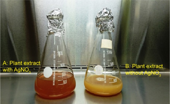
Materials and methods: We studied a green synthesis route to produce AgNPs by using an aqueous extract of Pimpinella anisum seeds (3 mM). Their antimicrobial activity and cytotoxicity on human neonatal skin stromal cells (hSSCs) and colon cancer cells (HT115) were assessed.
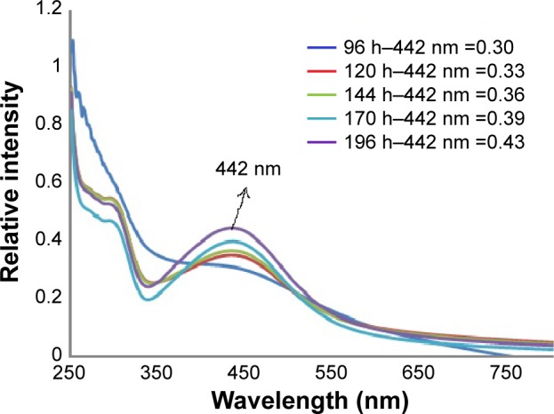
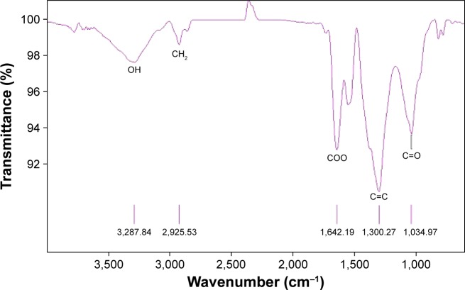
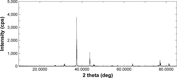
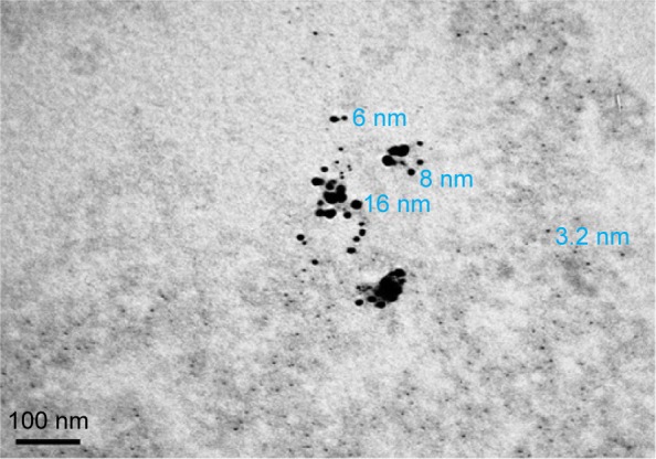
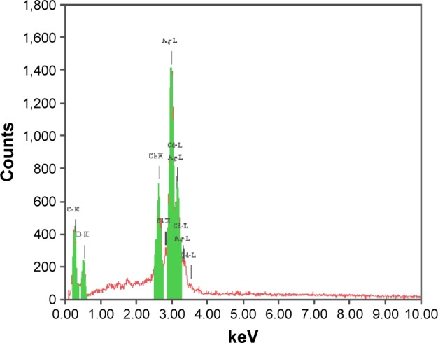
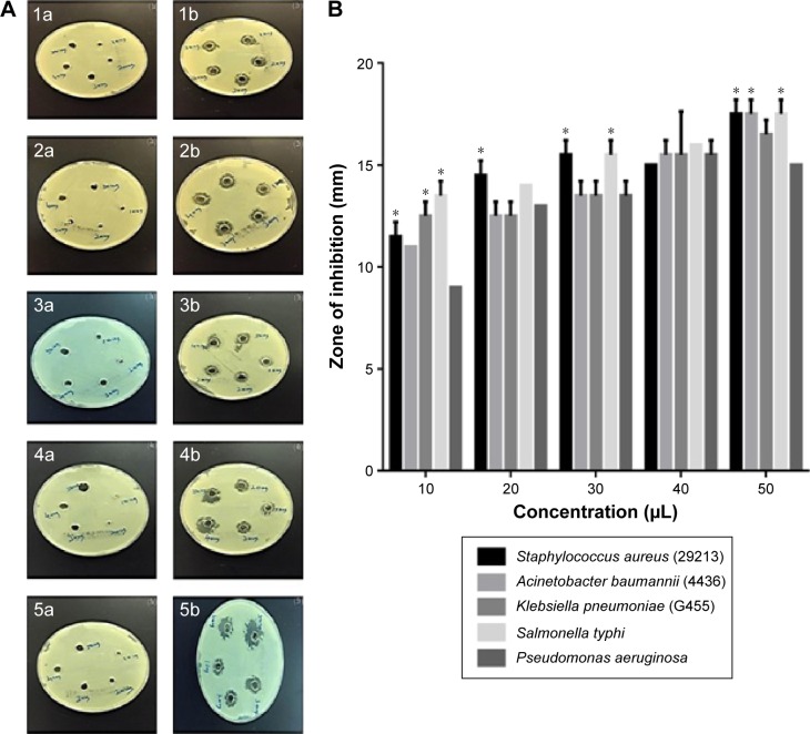
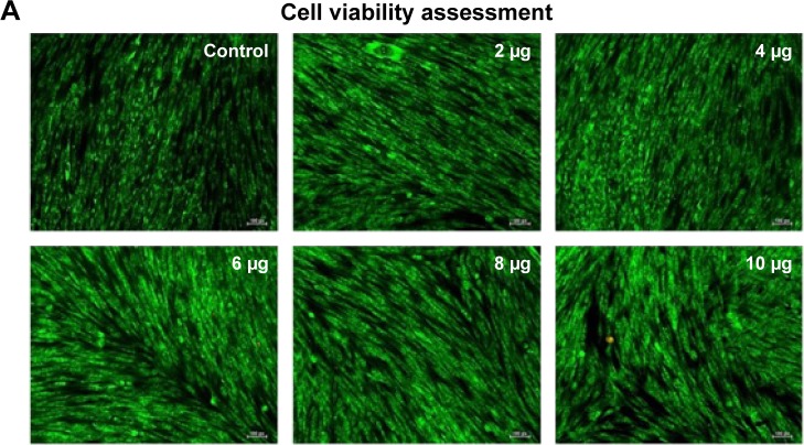
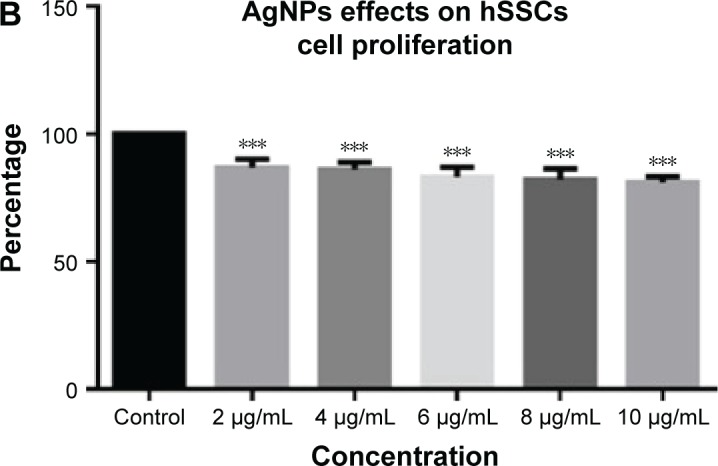
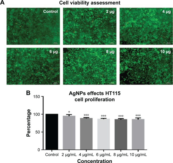
Results: A biophysical characterization of the synthesized AgNPs was realized: the morphology of AgNPs was determined by transmission electron microscopy, energy dispersive spectroscopy, X-ray powder diffraction, and ultraviolet-vis absorption spectroscopy. Transmission electron microscopy showed spherical shapes of AgNPs of P. anisum seed extracts with a 3.2 nm minimum diameter and average diameter ranging from 3.2 to 16 nm. X-ray powder diffraction highlighted the crystalline nature of the nanoparticles, ultraviolet-vis absorption spectroscopy was used to monitor their synthesis, and Fourier transform infrared spectroscopy showed the main reducing groups from the seed extract. Energy dispersive spectroscopy was used to confirm the presence of elemental silver. We evaluated the antimicrobial potential of green-synthesized AgNPs against five infectious bacteria: Staphylococcus pyogenes (29213), Acinetobacter baumannii (4436), Klebsiella pneumoniae (G455), Salmonella typhi, and Pseudomonas aeruginosa. In addition, we focused on the toxicological effects of AgNPs against hSSC cells and HT115 cells by using in vitro proliferation tests and cell viability assays. Among the different tested concentrations of nanoparticles, doses < 10 µg showed few adverse effects on cell proliferation without variations in viability, whereas doses >10 µg led to increased cytotoxicity.
Conclusion: Overall, our results highlighted the capacity of P. anisum-synthesized AgNPs as novel and cheap bioreducing agents for eco-friendly nanosynthetical routes. The data confirm the multipurpose potential of plant-borne reducing and stabilizing agents in nanotechnology.
Keywords: Pimpinella anisum seeds; antibacterial; biosafety; cancer; green nanotechnology; metal nanoparticles.
Figures
References
-
- Prashant KJ, Xiaohua H, Ivan HS, Mostafa AS. Noble metals on the nanoscale: optical and photothermal properties and some applications in imaging, sensing, biology, and medicine. Acc Chem Res. 2008;41(12):1578–1586. - PubMed
-
- Dahl JA, Maddux BL, Hutchison JE. Toward greener nanosynthesis. Chem Rev. 2007;107(6):2228–2269. - PubMed
-
- Raveendran P, Jie F, Scott LW. Completely “green” synthesis and stabilization of metal nanoparticles. J Am Chem Soc. 2003;125(46):13940–13941. - PubMed
-
- Benelli G. Plant-mediated biosynthesis of nanoparticles as an emerging tool against mosquitoes of medical and veterinary importance: a review. Parasitol Res. 2016;115(1):23–34. - PubMed
LinkOut - more resources
Full Text Sources
Other Literature Sources
