. 2016 Sep 8:7:1342.
doi: 10.3389/fmicb.2016.01342. eCollection 2016.

Leisingera sp. JC1, a Bacterial Isolate from Hawaiian Bobtail Squid Eggs, Produces Indigoidine and Differentially Inhibits Vibrios

Affiliations

Leisingera sp. JC1, a Bacterial Isolate from Hawaiian Bobtail Squid Eggs, Produces Indigoidine and Differentially Inhibits Vibrios

Samantha M Gromek et al. Front Microbiol. .

Abstract

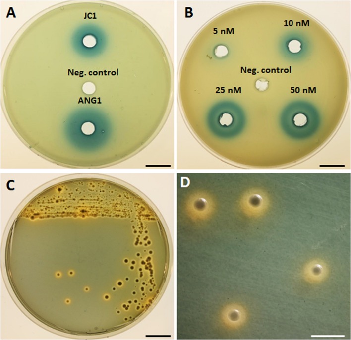
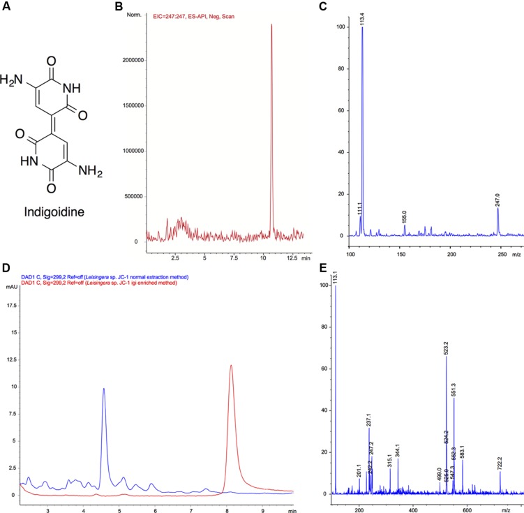
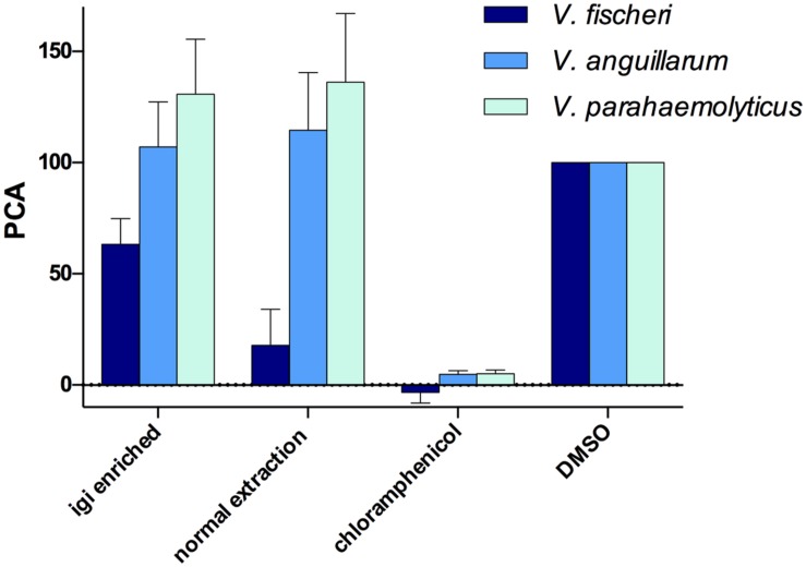
Female members of many cephalopod species house a bacterial consortium in the accessory nidamental gland (ANG), part of the reproductive system. These bacteria are deposited into eggs that are then laid in the environment where they must develop unprotected from predation, pathogens, and fouling. In this study, we characterized the genome and secondary metabolite production of Leisingera sp. JC1, a member of the roseobacter clade (Rhodobacteraceae) of Alphaproteobacteria isolated from the jelly coat of eggs from the Hawaiian bobtail squid, Euprymna scolopes. Whole genome sequencing and MLSA analysis revealed that Leisingera sp. JC1 falls within a group of roseobacters associated with squid ANGs. Genome and biochemical analyses revealed the potential for and production of a number of secondary metabolites, including siderophores and acyl-homoserine lactones involved with quorum sensing. The complete biosynthetic gene cluster for the pigment indigoidine was detected in the genome and mass spectrometry confirmed the production of this compound. Furthermore, we investigated the production of indigoidine under co-culture conditions with Vibrio fischeri, the light organ symbiont of E. scolopes, and with other vibrios. Finally, both Leisingera sp. JC1 and secondary metabolite extracts of this strain had differential antimicrobial activity against a number of marine vibrios, suggesting that Leisingera sp. JC1 may play a role in host defense against other marine bacteria either in the eggs and/or ANG. These data also suggest that indigoidine may be partially, but not wholly, responsible for the antimicrobial activity of this squid-associated bacterium.

Keywords: DART-MS; Euprymna; Leisingera; Rhodobacteraceae; indigoidine; roseobacter; secondary metabolite regulation; symbiosis.


Figures

FIGURE 1
FIGURE 1
MLSA analysis places Leisingera sp. JC1 in a sister group to other previously isolated ANG bacteria. Based on a comparison of 33 single-copy housekeeping genes, the egg jelly coat (JC) isolate JC1 is placed in the previously described roseobacter clade, “Clade 1” (Newton et al., 2010), along with seven previously isolated ANG bacteria (Collins et al., 2015). The phylogenetic tree reflects both Bayesian inference and maximum-likelihood methods with both posterior probability and bootstrap supports displayed.
FIGURE 2
FIGURE 2
Detection of homoserine lactone production and siderophore production by Leisingera sp. JC1. (A) Homoserine lactones were detected by β-galactosidase activity in cell-free supernatant of Leisingera sp. JC1 and compared with previously tested Leisingera sp. ANG1 (Collins et al., 2015); SWT broth was used as a negative control. (B) Dilution of N-3-oxohexanoyl homoserine lactone used as a positive control for the HSL assay; DMSO was the HSL standard solvent and was the negative control. (C) Leisingera sp. JC1 plated on CAS agar. Sequestration of iron changes the media from blue to orange, indicating siderophore production. (D) Magnified view of JC1 colonies from the plate in (C), showing the orange halos in the media indicative of siderophore production. Scale bars, 1.5 cm (A–C), 4 mm (D).
FIGURE 3
FIGURE 3
Leisingera sp. JC1 indigoidine biosynthesis operon. Operon organization obtained from the published genome sequence (OBY26161.1, OBY26149.1, OBY26162.1, OBY26150.1, OBY26151.1, OBY26152.1). Arrows drawn to scale.
FIGURE 4
FIGURE 4
Mass spectral confirmation of indigoidine production by Leisingera sp. JC1 and comparison of normal and igi enriched extracts. (A) Structure of indigoidine. (B) Extracted ion chromatogram (EIC) of indigoidine to confirm presence in extract ([M-H]- 247.0) using a previously reported LC–MS solvent gradient (Yu et al., 2013). (C) Negative ionization mass spectrum of peak at 10.7 min. The [M-H]- peak of 247.0 is consistent with a molecular formula of C10H7N4O4, confirming the presence of indigoidine (248.19 g/mol). (D) LC–MS UV chromatogram overlays (299 nm) of normal extract (blue) with indigoidine (igi) enriched extract (red). The peak eluting at tR 8.2 min in the igi enriched extract was confirmed to be indigoidine with a purity of 91.1% (tR different due to use of isocratic solvent system to minimize baseline variation). Although, a small peak was present at tR 8.2 min in the normal extract, this integrates to only 0.8% indicating that the Leisingera sp. JC1 normal extract contains negligible amounts of indigoidine. (E) Negative ionization mass spectrum of peak at tR 8.2 min for normal extraction method. Although there was an ion corresponding to [M-H]- of 247.2, this was not the most dominant ion and the more quantitative UV analysis in D shows only a very small peak, indicative of negligible indigoidine in the JC1 normal extract.
FIGURE 5
FIGURE 5
Leisingera sp. JC1 differentially inhibits pathogenic and non-pathogenic vibrio species. (A) Zones of inhibition around JC1 colonies were measured with increasing lawn densities of each vibrio species. Leisingera sp. JC1 differentially inhibited five vibrio strains. Significantly greater inhibition of Vibrio fischeri was seen between the 104–106 CFU/ml and the 107 CFU/ml lawn density. Significantly greater inhibition of Photobacterium leiognathi was seen between the 104 and 105 CFU/ml lawn densities, as well as between the 104–105 and the 106–107 CFU/ml densities. JC1 inhibited Vibrio anguillarum uniformly across all lawn densities and did not inhibit Vibrio parahaemolyticus or Vibrio harveyi. Letters indicate significantly different groups based on results from post hoc Tukey tests (see Results and Discussion). Representative 24 h images of (B) JC1 plated alone on YTSS agar and with 106 CFU/ml of (C) V. fischeri, (D) P. leiognathi, (E) V. anguillarum, (F) V. parahaemolyticus, and (G) V. harveyi. Scale bars: 5 mm (B–G).
FIGURE 6
FIGURE 6
Leisingera sp. JC1 extracts inhibit V. fischeri in 96-well liquid assays. JC1 cultures were grown at large scale and extracts were prepared using either an indigoidine enriched procedure (igi enriched) or a normal extraction method. All extracts were screened at 500 μg/mL against V. fischeri, V. anguillarum, and V. parahaemolyticus. The JC1 normal extract strongly inhibited growth of V. fischeri (PCA 17.9 ± 9.3) with less activity of the JC1 igi enriched extract against V. fischeri (PCA 63.3 ± 6.7). No inhibition of V. anguillarum or V. parahaemolyticus was observed. Data shown are average PCA values from three experiments (PCA, percent control activity, as compared with DMSO negative control).
FIGURE 7
FIGURE 7
Leisingera sp. JC1 localizes indigoidine production to outer edge of colony in the presence of V. fischeri. Five locations (A–E) were analyzed for the presence of indigoidine via direct analysis in real time-mass spectrometry (DART-MS) over 7 days. (A) In the absence of V. fischeri, indigoidine production was more uniformly distributed across the colony. (B) In the presence of V. fischeri, indigoidine production was primarily localized to outer edge of colony (location C) whereas minimal indigoidine was detected in the center of the colony (location A). Very little indigoidine was detected in the ZOI or outside ZOI in the presence or absence of V. fischeri. Heatmaps represent a gradient of indigoidine ion abundance from less abundance (black, 0%) to more abundance (red, 100%).
FIGURE 8
FIGURE 8
Leisingera sp. JC1 upregulates indigoidine production in the presence of V. fischeri. JC1 was cultured alone and in the presence of V. fischeri, V. anguillarum, and V. parahaemolyticus (each vibrio was also monocultured). (A) Extracts were prepared in DMSO at 5 mg/mL for LC–MS (from left to right): JC1 monoculture, JC1 + V. fischeri, JC1 + V. anguillarum, JC1 + V. parahaemolyticus, V. fischeri monoculture, V. anguillarum monoculture, and V. parahaemolyticus monoculture. As shown, the highest pigment production was observed when JC1 was co-cultured with V. fischeri. (B) LC–MS UV chromatograms (299 nm) for all extracts. JC1 indigoidine production was upregulated when co-cultured with V. fischeri (red). Down regulation was observed when co-cultured with V. anguillarum or V. parahaemolyticus (green and pink, respectively). (C) Following extraction, indigoidine production was measured using HPLC and quantitated via measurement of the area under the curve for UV absorption at 299 nm. Fold change was calculated as compared with JC1 monoculture.

References

    1. Alikhan N.-F., Petty N. K., Ben Zakour N. L., Beatson S. A. (2011). BLAST Ring Image Generator (BRIG): simple prokaryote genome comparisons. BMC Genomics 12:402 10.1186/1471-2164-12-402 - DOI - PMC - PubMed
    1. Amsler C. D., McClintock J. B., Baker B. J. (2001). Secondary metabolites as mediators of trophic interactions among Antarctic marine organisms. Am. Zool. 41 17–26.
    1. Antonic V., Stojadinovic A., Zhang B., Izadjoo M. J., Alavi M. (2013). Pseudomonas aeruginosa induces pigment production and enhances virulence in a white phenotypic variant of Staphylococcus aureus. Infect. Drug Resist. 6 175–186. 10.2147/IDR.S49039 - DOI - PMC - PubMed
    1. Aziz R. K., Bartels D., Best A. A., DeJongh M., Disz T., Edwards R. A., et al. (2008). The RAST Server: rapid annotations using subsystems technology. BMC Genomics 9:75 10.1186/1471-2164-9-75 - DOI - PMC - PubMed
    1. Barbieri E., Barry K., Child A., Wainwright N. (1997). Antimicrobial activity in the microbial community of the accessory nidamental gland and egg cases of Loligo pealei (Cephalopoda: Loliginidae). Biol. Bull. 193 275–276. - PubMed

LinkOut - more resources