. 2016 Sep 20:4:55.
doi: 10.1186/s40425-016-0157-6. eCollection 2016.

A case report of Grover's disease from immunotherapy-a skin toxicity induced by inhibition of CTLA-4 but not PD-1

Affiliations

A case report of Grover's disease from immunotherapy-a skin toxicity induced by inhibition of CTLA-4 but not PD-1

Marc Uemura et al. J Immunother Cancer. .

Erratum in

Abstract

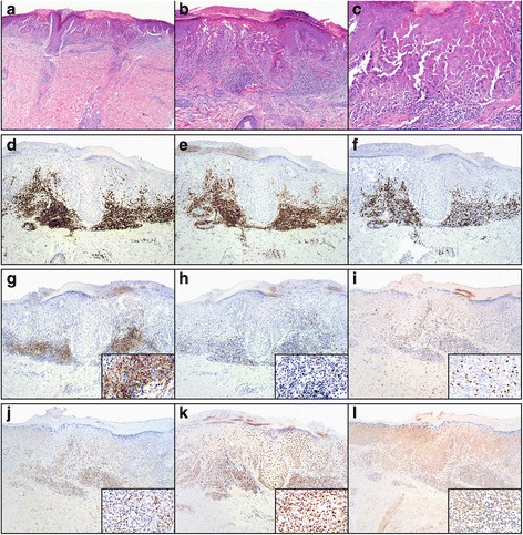
Background: Immune related adverse events (irAEs) are common side effects of checkpoint inhibitory (CPI) therapies targeting CTLA-4 and PD-1/PD-L1. Grover's disease is an uncommon dermatologic condition with unclear pathogenesis previously reported as an irAE with ipilimumab.

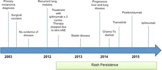
Case presentation: We report an additional case of ipilimumab-induced Grover's disease. Interestingly, this dermatologic side effect did not appear with use of anti-PD-1 therapy in our patient. Immune analysis was performed and suggests a possible role of Th2 cells in its patholgenesis.

Conclusion: This case suggests that Grover's disease is an irAE induced by Ipilimumab. Our immune analysis suggests that Th2 cells may be pathogenic mediators which warrants further study.


Figures

Fig. 1
Fig. 1
Histopathologic and immunophenotypic studies of ipilimumab-induced Grover’s disease. a-c Scanning magnification of skin with acantholytic dyskeratosis (a, H&E, 20×). Higher magnification reveals skin with suprabasal acantholysis and overlying dyskeratosis; (b, H&E, 100×) and (c, H&E, 40×). Immunohistochemical studies demonstrate a predominance of CD3+ T-cells (d, anti-CD3, 100×) comprised of a predominance of CD4+ T-cells (e, anti-CD4, 100×) over CD8+ T-cells (f, anti-CD8, 100×). Additional immunohistochemical studies demonstrate strong expression of PD-L1 by the inflammatory cells (g, anti-PD-L1, 100×; inset, 400×). Scattered cells express PD-1 (h, anti-PD-1, 100×; inset 400×). There is scattered nuclear expression of FoxP3 (i, anti-FoxP3, 100×; inset 400×) and T-beta (j, anti-T-Bet, 100×; inset, 400×) but strong diffuse nuclear expression of Gata-3 by the majority of the inflammatory cells as well as the overlying keratinocytes (k, anti-Gata-3, 100×; inset: 400×). Antibodies for RORgT are essentially negative in the infiltrate. There is strong background staining in the skin tissue (l, anti-RORgT, 100×; inset: 400×)
Fig. 2
Fig. 2
Chronologic timeline of patient’s clinical course

References

    1. Grover RW. Transient acantholytic dermatosis. Arch Dermatol. 1970;101(4):426–434. doi: 10.1001/archderm.1970.04000040048010. - DOI - PubMed
    1. Chalet M, Grover R, Ackerman AB. Transient acantholytic dermatosis: a reevaluation. Arch Dermatol. 1977;113(4):431–435. doi: 10.1001/archderm.1977.01640040039004. - DOI - PubMed
    1. Davis MD, Dinneen AM, Landa N, Gibson LE. Grover's disease: clinicopathologic review of 72 cases. Mayo Clin Proc. 1999;74(3):229–234. doi: 10.4065/74.3.229. - DOI - PubMed
    1. Guana AL, Cohen PR. Transient acantholytic dermatosis in oncology patients. J Clin Oncol. 1994;12(8):1703–1709. - PubMed
    1. Anforth R, Fernandez-Penas P, Long GV. Cutaneous toxicities of RAF inhibitors. Lancet Oncol. 2013;14(1):e11–18. doi: 10.1016/S1470-2045(12)70413-8. - DOI - PubMed

LinkOut - more resources