. 2016 Sep 23;11(9):e0163645.
doi: 10.1371/journal.pone.0163645. eCollection 2016.

Increased Brain-Specific MiR-9 and MiR-124 in the Serum Exosomes of Acute Ischemic Stroke Patients

Affiliations

Increased Brain-Specific MiR-9 and MiR-124 in the Serum Exosomes of Acute Ischemic Stroke Patients

Qiuhong Ji et al. PLoS One. .

Abstract

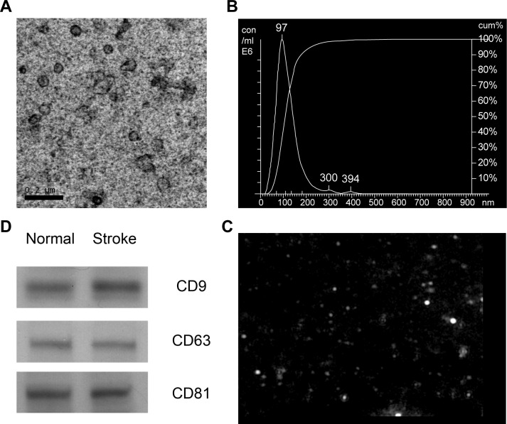
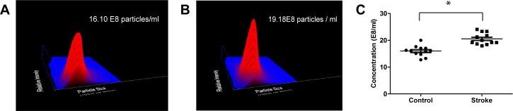
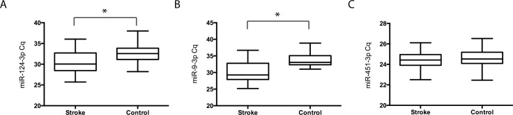
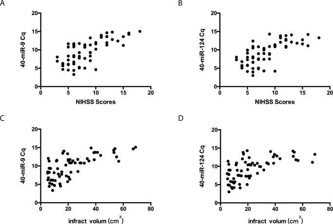
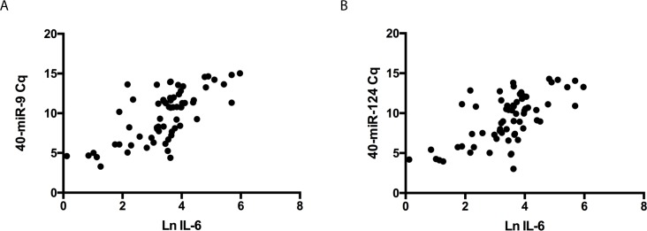
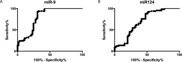
The aims of this study were to examine the alternation in serum exosome concentrations and the levels of serum exosomal miR-9 and miR-124, two brain-specific miRNAs, in acute ischemic stroke (AIS) patients and to explore the predictive values of these miRNAs for AIS diagnosis and damage evaluation. Sixty-five patients with AIS at the acute stage were enrolled and 66 non-stroke volunteers served as controls. Serum exosomes isolated by ExoQuick precipitations were characterized by transmission electron microscopy, nanoparticle-tracking analysis and western blotting. The levels of exosomal miR-9 and miR-124 were determined by real-time quantitative PCR. Compared with controls, the concentration of serum exosomes and the median levels of serum exosomal miR-9 and miR-124 were significantly higher in AIS patients (p<0.01). The levels of both miR-9 and miR-124 were positively correlated with National Institutes of Health Stroke Scale (NIHSS) scores, infarct volumes and serum concentrations of IL-6. The areas under the curve for exosomal miR-9 and miR-124 were 0.8026 and 0.6976, respectively. This proof of concept study suggests that serum exosomal miR-9 and miR-124 are promising biomarkers for diagnosing AIS and evaluating the degree of damage caused by ischemic injury. However, further studies are needed to explore the potential roles of the exosomes released from brain tissues in post stroke complications.


Conflict of interest statement

The authors have declared that no competing interests exist.

Figures

Fig 1
Fig 1. Characterization of isolated exosomes from human serum.
(A) The TEM image shows the spherical morphology of exosomes with a diameter of approximately 100 nm, bar = 200 nm. Representative images show the size distribution (B) and morphology (C) of serum exosomes measured by NTA. (D) Western blotting analysis of exosome markers, CD9, CD63 and CD81, in the serum exosomes from non-stroke controls and stroke patients.
Fig 2
Fig 2. Comparison of serum exosomal concentrations of non-stroke controls and the stroke group.
Representative plots of NTA analysis of serum exosomes from a non-stroke individual (A) and an AIS patient (B). The indicated concentration is the value for 1,000 diluted samples. (C). Statistical analysis of the serum exosomal concentrations of AIS patients and non-stroke controls, *, p<0.01.
Fig 3
Fig 3. Comparison of levels of serum exosomal miR-9, miR-124 and miR-451 between stroke patients and non-stroke controls.
Relative serum exosomal miR-9 (A), miR-124 (B) and miR-451 (C) levels between the stroke group (n = 65) and non-stroke control groups (n = 66). The y-axis depicts Cq values normalized to cel-miR-39. The black horizontal line in the box represents the median value of the group. *, p<0.01.
Fig 4
Fig 4
Correlations between the levels of serum exosomal miR-9 (A), miR-124 (B) and the NIHSS score in 65 AIS patients at an acute stage. Correlations between the levels of serum exosomal miR-9 (C), miR-124 (D) and the infarction volume in 65 stroke patients at an acute stage. The scatter plots show significant positive correlations between the serum exosomal miR-9 and miR-124 levels (expressed as 40-Delta Cq) and NIHSS scores and infarct volumes.
Fig 5
Fig 5
Correlations between the levels of serum exosomal miR-9 (A) and miR-124 (B) and the serum concentrations of IL-6 in stroke patients. The scatter plots show significant positive correlations between the serum exosomal miR-9 and miR-124 levels (expressed as 40-Delta Cq) and Ln transferred IL-6 concentrations.
Fig 6
Fig 6. Diagnostic value of serum exosomal miR-9 and miR-124 levels in AIS.
ROC curves based on exosomal miR-9 (A) and miR-124 (B) levels to differentiate AIS patients (n = 65) from non-stroke individuals (n = 66). The areas under the curve (AUC) are 0.8026 (95% CI: 0.7235–0.8816) and 0.6976 (95% CI: 0.6506–0.7895).

References

    1. Jickling GC, Sharp FR. Biomarker panels in ischemic stroke. Stroke; a journal of cerebral circulation. 2015;46(3):915–20. 10.1161/STROKEAHA.114.005604 - DOI - PMC - PubMed
    1. Reichlin T, Hochholzer W, Bassetti S, Steuer S, Stelzig C, Hartwiger S, et al. Early diagnosis of myocardial infarction with sensitive cardiac troponin assays. The New England journal of medicine. 2009;361(9):858–67. 10.1056/NEJMoa0900428 . - DOI - PubMed
    1. Bartel DP. MicroRNAs: genomics, biogenesis, mechanism, and function. Cell. 2004;116(2):281–97. . - PubMed
    1. Lagos-Quintana M, Rauhut R, Yalcin A, Meyer J, Lendeckel W, Tuschl T. Identification of tissue-specific microRNAs from mouse. Current biology: CB. 2002;12(9):735–9. . - PubMed
    1. Weber JA, Baxter DH, Zhang S, Huang DY, Huang KH, Lee MJ, et al. The microRNA spectrum in 12 body fluids. Clinical chemistry. 2010;56(11):1733–41. 10.1373/clinchem.2010.147405 . - DOI - PMC - PubMed