. 2016 Oct 11;7(41):66444-66454.
doi: 10.18632/oncotarget.12119.

β-Hydroxybutyrate suppresses inflammasome formation by ameliorating endoplasmic reticulum stress via AMPK activation

Affiliations

β-Hydroxybutyrate suppresses inflammasome formation by ameliorating endoplasmic reticulum stress via AMPK activation

Ha Ram Bae et al. Oncotarget. .

Abstract

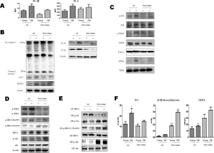
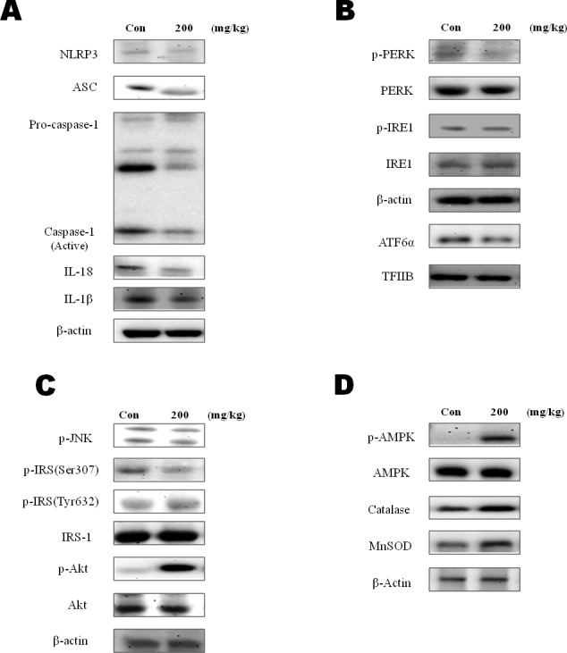
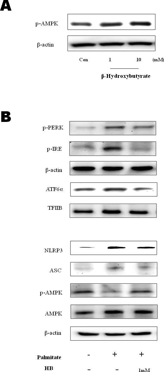
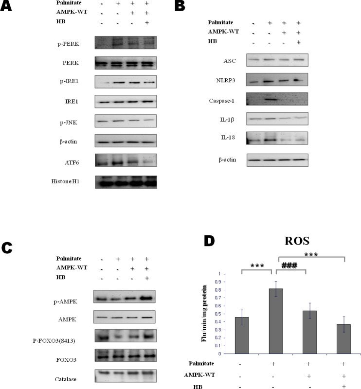
β-Hydroxybutyrate, a ketone body that is used as an energy source in organs such as the brain, muscle, and heart when blood glucose is low, is produced by fatty acid oxidation in the liver under the fasting state. Endoplasmic reticulum (ER) stress is linked with the generation of intracellular reactive oxygen species and the accumulation of misfolded protein in the ER. ER stress is known to induce the NOD-like receptor protein 3 inflammasome, which mediates activation of the proinflammatory cytokine interleukin-1β, whose maturation is caspase-1-dependent. We investigated whether β-hydroxybutyrate modulates ER stress, inflammasome formation, and insulin signaling. Sprague Dawley rats (6 and 24 months of age) that were starved for 3 d and rats treated with β-hydroxybutyrate (200 mg·kg-1·d-1 i.p., for 5 d) were used for in vivo investigations, whereas human hepatoma HepG2 cells were used for in vitro studies. Overexpression of AMPK in cultured cells was performed to elucidate the molecular mechanism. The starvation resulted in increased serum β-hydroxybutyrate levels with decreased ER stress (PERK, IRE1, and ATF6α) and inflammasome (ASC, caspase-1, and NLRP3) formation compared with non-fasted 24-month-old rats. In addition, β-hydroxybutyrate suppressed the increase of ER stress- and inflammasome-related marker proteins. Furthermore, β-hydroxybutyrate treatment increased the expression of manganese superoxide dismutase and catalase via the AMP-activated protein kinase-forkhead box protein O3α transcription factor pathway both in vivo and in vitro. The significance of the current study was the discovery of the potential therapeutic role of β-hydroxybutyrate in suppressing ER-stress-induced inflammasome formation.

Keywords: AMPK; Gerotarget; aging; endoplasmic reticulum; inflammasome; β-hydroxybutyrate.


Conflict of interest statement

CONFLICTS OF INTEREST

The authors declare no conflict of interest.

Figures

Figure 1
Figure 1. Modulation of ER-stress-induced inflammasome formation in acute starvation rat liver
A. IL-1β and IL-6 levels were determined in the serum. Results of one-factor ANOVA: *p < 0.05 vs. Ad libitum. B. Western blot analysis was performed to determine the inflammasome levels in the cytoplasmic extracts. As shown, the protein levels of the inflammasome were decreased by acute starvation. C. Western blot analysis was performed to determine the protein levels of UPR markers. As shown, the levels were significantly decreased in the starvation model. One representative blot of each protein is shown from three experiments that yielded similar results. D. Western blot analysis was performed to detect the presence of insulin signaling in the cytoplasmic extracts. The protein levels of serine phosphorylation of IRS-1 and phosphorylation of Akt were decreased by starvation. In contrast, tyrosine phosphorylation of IRS-1 was increased. E. IRS-2 interaction of Akt and its modulation by starvation. cytosol extracts were prepared from young and old rat livers. Immunoprecipitated IRS-2 was determined to be physically associated with p-seine, p-tyrosine, p-IRS-1/2 (t632), p-Akt, and Akt by Western blotting. F. Constituents were determined in the serum. Results of one-factor ANOVA: *p < 0.05 vs. Ad libitum.
Figure 2
Figure 2. Modulation of β-hydroxybutyrate on ER-stress-induced inflammasome formation and the AMPK pathway
A. Western blot analysis was performed to detect the presence of caspase-1, ASC, NLRP3, β-actin, IL-18, and IL-1β in liver homogenates from β-hydroxybutyrate-treated rats (200 mg·kg−1·day−1 for 5 d). B. To determine ER stress marker changes by β-hydroxybutyrate treatment, the expressions of p-PERK, p-IRE1, and ATF6α were examined. C. Western blot analysis was performed to detect the presence of phosphorylated IRS-1 at Ser 307 and Tyr 632, phosphorylated Akt, and phosphorylated JNK in liver homogenates. D. Western blot analysis was performed to detect the presence of p-AMPK, catalase, and SOD-2 (MnSOD) in liver homogenates.
Figure 3
Figure 3. Effect of β-hydroxybutyrate on AMPK activation and ER-stress-induced inflammasome formation in HepG2 cells
A. HepG2 cells were treated with β-hydroxybutyrate (1 and 10 mM). Cells were lysed after 2 h of treatment, and western blot analysis was performed to determine the change of phosphorylation of AMPK in the cytosolic fraction. B. HepG2 cells were treated with 1 mM β-hydroxybutyrate and incubated. After 2 h, the cells were treated with 250 μM palmitate. Levels of p-PERK, p-IRE1, ATF6α, NLRP3, ASC, p-AMPK, and AMPK were then measured by western blot analysis using their specific antibodies. β-Actin and TFIIB blot analyses are shown, to clarify the same amount of proteins loaded in the cytosolic and nuclear fractions, respectively.
Figure 4
Figure 4. Effect of β-hydroxybutyrate on ER-stress-induced inflammasome formation via the AMPK pathway in vitro
A. HepG2 cells were transfected with lentiviral particle wild-type AMPK. After 12 h, cells were treated with 1 mM β-hydroxybutyrate, followed by 250 μM palmitate for 1 h. Western blot analysis was performed to determine the levels of p-PERK, p-IRE1, p-JNK, and ATF6α. β-Actin and histone H1 blots are shown, to clarify the same amount of protein loaded in the cytosolic and nuclear fractions, respectively. B., C. HepG2 cells were transfected with lentiviral particle wild-type AMPK. After 12 h, cells were treated with 1 mM β-hydroxybutyrate, followed by 250 μM palmitate for 1 h. D. Reactive oxygen species generation was measured by dichlorofluorescein formation with the fluorescent probe 2′,7′-dichlorofluorescein diacetate. Results of one-factor ANOVA: ***p < 0.001 vs. control; ###p < 0.001 vs. palmitate. HB, β-hydroxybutyrate.

References

    1. Veech RL, Chance B, Kashiwaya Y, Lardy HA, Cahill GF., Jr Ketone bodies, potential therapeutic uses. IUBMB Life. 2001;51:241–247. - PubMed
    1. Cahill GF, Jr, Herrera MG, Morgan AP, Soeldner JS, Steinke J, Levy PL, Reichard GA, Jr, Kipnis DM. Hormone-fuel interrelationships during fasting. J Clin Invest. 1996;45:1751–1769. - PMC - PubMed
    1. Laffel L. Ketone bodies: a review of physiology, pathophysiology and application of monitoring to diabetes. Diabetes Metab Res Rev. 1999;15:412–426. - PubMed
    1. Hegardt FG. Mitochondrial 3-hydroxy-3-methylglutaryl-CoA synthase : a control enzyme in ketogenesis. Biochem J. 1999;338:569–582. - PMC - PubMed
    1. Owen OE, Felig P, Morgan AP, Wahren J, Cahill GF., Jr Liver and kidney metabolism during prolonged starvation. J Clin Invest. 1969;48:574–583. - PMC - PubMed