

TWIST1/miR-584/TUSC2 pathway induces resistance to apoptosis in thyroid cancer cells
- PMID: 27661106
- PMCID: PMC5342575
- DOI: 10.18632/oncotarget.12129
TWIST1/miR-584/TUSC2 pathway induces resistance to apoptosis in thyroid cancer cells
Abstract
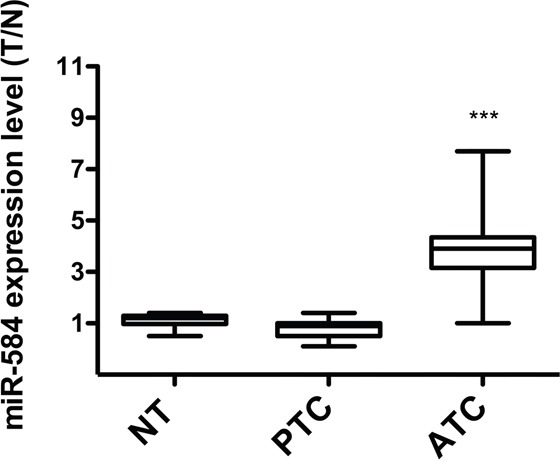
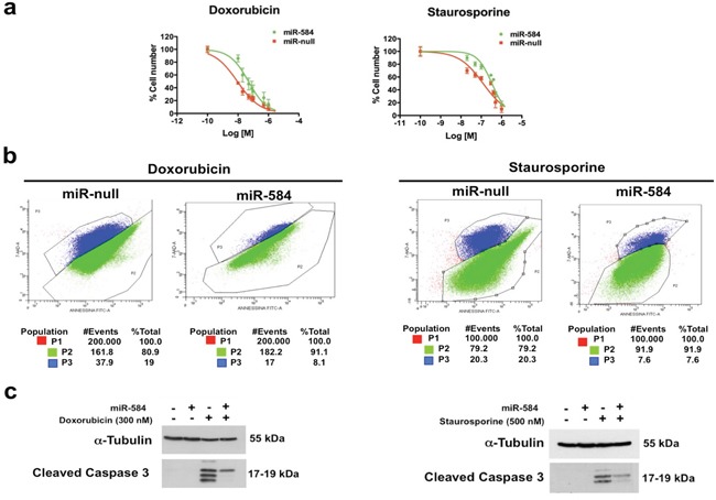
TWIST1, a transcription factor, plays a pivotal role in cancer initiation and progression. Anaplastic thyroid carcinoma (ATC) is one of the deadliest human malignancies; TWIST1 is overexpressed in ATC and increases thyroid cancer cell survival, migration and invasion. The molecular mechanisms underlying the effects of TWIST1 are partially known. Here, using miRNome profiling of papillary thyroid cancer cells (TPC-1) ectopically expressing TWIST1, we identified miR-584. We showed that TWIST1 directly binds miR-584 using chromatin immunoprecipitation. Importantly, miR-584 was up-regulated in human ATC compared to papillary thyroid carcinoma (PTC) and normal thyroid samples. Overexpression of miR-584 in TPC cells induced resistance to apoptosis, whereas stable transfection of anti-miR-584 in TPC-TWIST1 and 8505C cells increased the sensitivity to apoptosis. Using bioinformatics programs, we identified TUSC2 (tumor suppressor candidate 2) as a novel target of miR-584. TUSC2 mRNA and protein levels were decreased in TPC miR-584 and increased in TPC-TWIST1 anti-miR-584 cells. Luciferase assays demonstrated direct targeting. Restored expression of TUSC2 rescued the inhibition of apoptosis induced by miR-584. Finally, qRT-PCR and immunohistochemical analysis showed that TUSC2 was down-regulated in ATC and PTC samples compared to normal thyroids. In conclusion, our study identified a novel TWIST1/miR-584/TUSC2 pathway that plays a role in resistance to apoptosis of thyroid cancer cells.
Keywords: TUSC2; TWIST1; anaplastic thyroid carcinoma; apoptosis; miR-584.
Conflict of interest statement
The authors declare no conflicts of interest.
Figures


Similar articles
-
Identification of targets of Twist1 transcription factor in thyroid cancer cells.J Clin Endocrinol Metab. 2014 Sep;99(9):E1617-26. doi: 10.1210/jc.2013-3799. Epub 2014 May 21. J Clin Endocrinol Metab. 2014. PMID: 24848707
-
The TUSC2 Tumour Suppressor Inhibits the Malignant Phenotype of Human Thyroid Cancer Cells via SMAC/DIABLO Protein.Int J Mol Sci. 2020 Jan 21;21(3):702. doi: 10.3390/ijms21030702. Int J Mol Sci. 2020. PMID: 31973107 Free PMC article.
-
MiR-20b Displays Tumor-Suppressor Functions in Papillary Thyroid Carcinoma by Regulating the MAPK/ERK Signaling Pathway.Thyroid. 2016 Dec;26(12):1733-1743. doi: 10.1089/thy.2015.0578. Epub 2016 Nov 2. Thyroid. 2016. PMID: 27717302
-
Expression of MicroRNAs in Thyroid Carcinoma.Methods Mol Biol. 2017;1617:261-280. doi: 10.1007/978-1-4939-7046-9_19. Methods Mol Biol. 2017. PMID: 28540691 Review.
-
miR-451a is underexpressed and targets AKT/mTOR pathway in papillary thyroid carcinoma.Oncotarget. 2016 Mar 15;7(11):12731-47. doi: 10.18632/oncotarget.7262. Oncotarget. 2016. PMID: 26871295 Free PMC article. Review.
Cited by
-
Tumor suppressor candidate 2 (TUSC2, FUS-1) and human cancers.Discov Med. 2017 May;23(128):325-330. Discov Med. 2017. PMID: 28715648 Free PMC article. Review.
-
Immunohistochemical and molecular evaluation of TUSC2 expression in breast cancer.Mol Biol Rep. 2024 Mar 6;51(1):394. doi: 10.1007/s11033-024-09320-z. Mol Biol Rep. 2024. PMID: 38446366
-
Clinical Study of Virtual Reality Augmented Technology Combined with Contrast-Enhanced Ultrasound in the Assessment of Thyroid Cancer.J Healthc Eng. 2021 Aug 6;2021:8042755. doi: 10.1155/2021/8042755. eCollection 2021. J Healthc Eng. 2021. Retraction in: J Healthc Eng. 2023 Jun 21;2023:9861340. doi: 10.1155/2023/9861340. PMID: 34394897 Free PMC article. Retracted.
-
miR-650 promotes motility of anaplastic thyroid cancer cells by targeting PPP2CA.Endocrine. 2019 Sep;65(3):582-594. doi: 10.1007/s12020-019-01910-3. Epub 2019 Mar 29. Endocrine. 2019. PMID: 30927143
-
MicroRNA-584 inhibits cell proliferation and invasion in non-small cell lung cancer by directly targeting MTDH.Exp Ther Med. 2018 Feb;15(2):2203-2211. doi: 10.3892/etm.2017.5624. Epub 2017 Dec 12. Exp Ther Med. 2018. Retraction in: Exp Ther Med. 2022 Jun 14;24(2):515. doi: 10.3892/etm.2022.11442. PMID: 29434826 Free PMC article. Retracted.
References
-
- Ansieau S, Morel AP, Hinkal G, Bastid J, Puisieux A. TWISTing an embryonic transcription factor into an oncoprotein. Oncogene. 2010;29:3173–3184. - PubMed
-
- Aparicio LA, Blanco M, Castosa R, Concha A, Valladares M, Calvo L, Figueroa A. Clinical implications of epithelial cell plasticity in cancer progression. Cancer letters. 2015;366:1–10. - PubMed
MeSH terms
Substances
LinkOut - more resources
Full Text Sources
Other Literature Sources
Medical
Molecular Biology Databases
Research Materials
