Acquired resistance to venetoclax (ABT-199) in t(14;18) positive lymphoma cells
- PMID: 27661108
- PMCID: PMC5342530
- DOI: 10.18632/oncotarget.12132
Acquired resistance to venetoclax (ABT-199) in t(14;18) positive lymphoma cells
Abstract
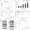
The chromosomal translocation t(14;18) in follicular lymphoma (FL) is a primary oncogenic event resulting in BCL-2 over-expression. This study investigates activity of the BH3 mimetic venetoclax (ABT-199), which targets BCL-2, and mechanisms of acquired resistance in FL.The sensitivity of FL cells to venetoclax treatment correlated with BCL-2/BIM ratio. Cells with similar expression of anti-apoptotic proteins, but with higher levels of BIM were more sensitive to the treatment. Venetoclax induced dissociation of BCL-2/ BIM complex and a decrease in mitochondrial potential. Interestingly the population of cells that survived venetoclax treatment showed increased p-ERK1/2 and p-BIM (S69), as well as a decrease in total BIM levels. Venetoclax resistant cells initially showed elevated levels of p-AKT and p-Foxo1/3a, a dissociation of BIM/BCL-2/BECLIN1 complex, and a decrease in SQSTM1/p62 level (indicating increased autophagy) together with a slight decline in BIM expression. After stable resistant cell lines were established, a significant reduction of BCL-2 levels and almost total absence of BIM was observed.The acquisition of these resistance phenotypes could be prevented via selective ERK/AKT inhibition or anti-CD20 antibody treatment, thus highlighting possible combination therapies for FL patients.
Keywords: follicular lymphoma; resistance; venetoclax.
Conflict of interest statement
None.
Figures


Similar articles
-
Differential Expression of Bcl-2 Family Proteins Determines the Sensitivity of Human Follicular Lymphoma Cells to Dexamethasone-mediated and Anti-BCR-mediated Apoptosis.J Immunother. 2016 Jan;39(1):8-14. doi: 10.1097/CJI.0000000000000102. J Immunother. 2016. PMID: 26641257
-
A synthetic peptide targeting the BH4 domain of Bcl-2 induces apoptosis in multiple myeloma and follicular lymphoma cells alone or in combination with agents targeting the BH3-binding pocket of Bcl-2.Oncotarget. 2015 Sep 29;6(29):27388-402. doi: 10.18632/oncotarget.4489. Oncotarget. 2015. PMID: 26317541 Free PMC article.
-
Potential mechanisms of resistance to venetoclax and strategies to circumvent it.BMC Cancer. 2017 Jun 2;17(1):399. doi: 10.1186/s12885-017-3383-5. BMC Cancer. 2017. PMID: 28578655 Free PMC article.
-
Targeting Bcl-2 for the treatment of multiple myeloma.Leukemia. 2018 Sep;32(9):1899-1907. doi: 10.1038/s41375-018-0223-9. Epub 2018 Aug 3. Leukemia. 2018. PMID: 30076373 Review.
-
Therapeutics targeting Bcl-2 in hematological malignancies.Biochem J. 2017 Oct 23;474(21):3643-3657. doi: 10.1042/BCJ20170080. Biochem J. 2017. PMID: 29061914 Review.
Cited by
-
A novel CDK9 inhibitor increases the efficacy of venetoclax (ABT-199) in multiple models of hematologic malignancies.Leukemia. 2020 Jun;34(6):1646-1657. doi: 10.1038/s41375-019-0652-0. Epub 2019 Dec 11. Leukemia. 2020. PMID: 31827241 Free PMC article.
-
Restoring Apoptosis with BH3 Mimetics in Mature B-Cell Malignancies.Cells. 2020 Mar 14;9(3):717. doi: 10.3390/cells9030717. Cells. 2020. PMID: 32183335 Free PMC article. Review.
-
BH3 Mimetics for the Treatment of B-Cell Malignancies-Insights and Lessons from the Clinic.Cancers (Basel). 2020 Nov 12;12(11):3353. doi: 10.3390/cancers12113353. Cancers (Basel). 2020. PMID: 33198338 Free PMC article. Review.
-
Programmed cell death, redox imbalance, and cancer therapeutics.Apoptosis. 2021 Aug;26(7-8):385-414. doi: 10.1007/s10495-021-01682-0. Epub 2021 Jul 8. Apoptosis. 2021. PMID: 34236569 Review.
-
Synergistic Action of MCL-1 Inhibitor with BCL-2/BCL-XL or MAPK Pathway Inhibitors Enhances Acute Myeloid Leukemia Cell Apoptosis and Differentiation.Int J Mol Sci. 2023 Apr 13;24(8):7180. doi: 10.3390/ijms24087180. Int J Mol Sci. 2023. PMID: 37108344 Free PMC article.
References
-
- Gupta RK, Lister TA. Current management of follicular lymphoma. Curr Opin Oncol. 1996;8:360–365. - PubMed
-
- Tsujimoto Y, Cossman J, Jaffe E, Croce CM. Involvement of the bcl-2 gene in human follicular lymphoma. Science. 1985;228:1440–1443. - PubMed
-
- Roberts AW, Seymour JF, Brown JR, Wierda WG, Kipps TJ, Khaw SL, Carney DA, He SZ, Huang DC, Xiong H, Cui Y, Busman TA, McKeegan EM, et al. Substantial susceptibility of chronic lymphocytic leukemia to BCL2 inhibition: results of a phase I study of navitoclax in patients with relapsed or refractory disease. J Clin Oncol. 2012;30:488–496. - PMC - PubMed
MeSH terms
Substances
LinkOut - more resources
Full Text Sources
Other Literature Sources
Medical
Molecular Biology Databases
Research Materials
Miscellaneous

