. 2016 Oct 25;7(43):70194-70210.
doi: 10.18632/oncotarget.12169.

MicroRNA-552 enhances metastatic capacity of colorectal cancer cells by targeting a disintegrin and metalloprotease 28

Affiliations

MicroRNA-552 enhances metastatic capacity of colorectal cancer cells by targeting a disintegrin and metalloprotease 28

Jian Wang et al. Oncotarget. .

Abstract

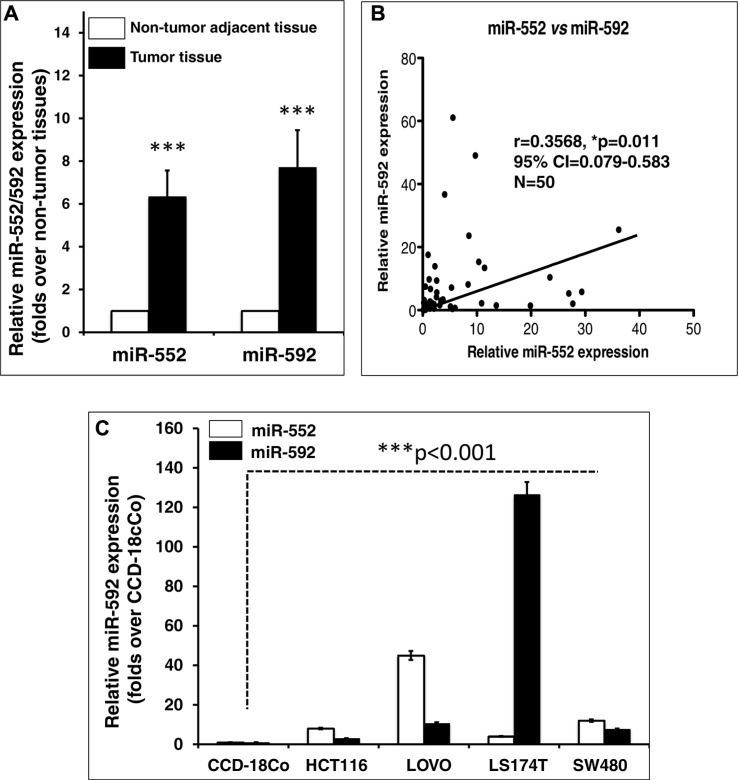
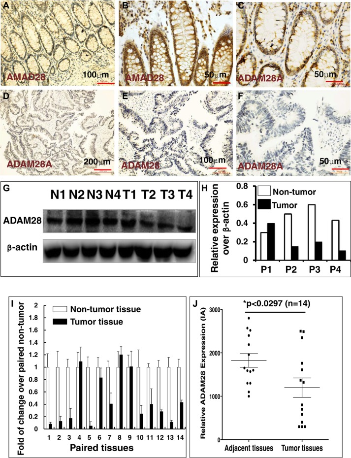
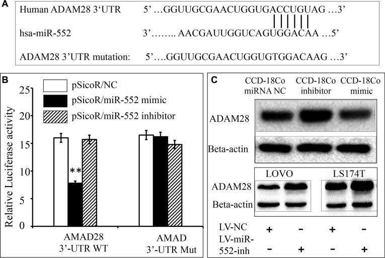
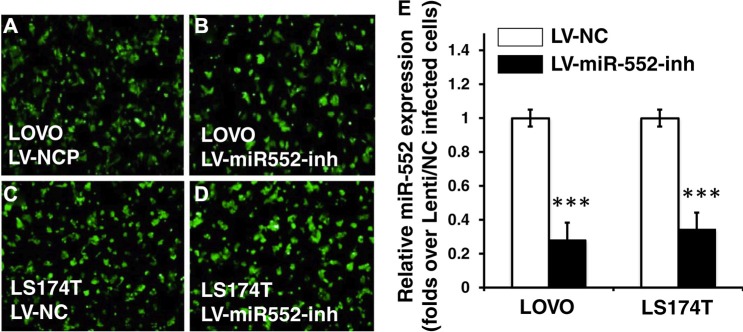
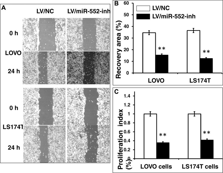
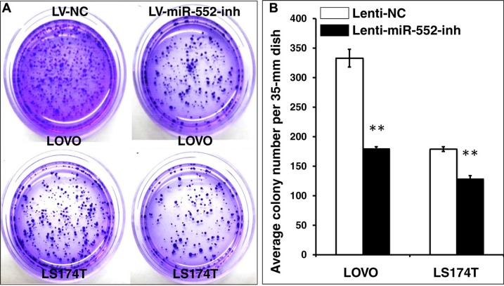
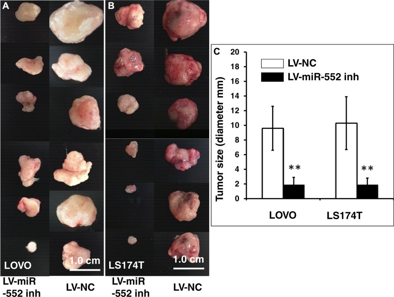
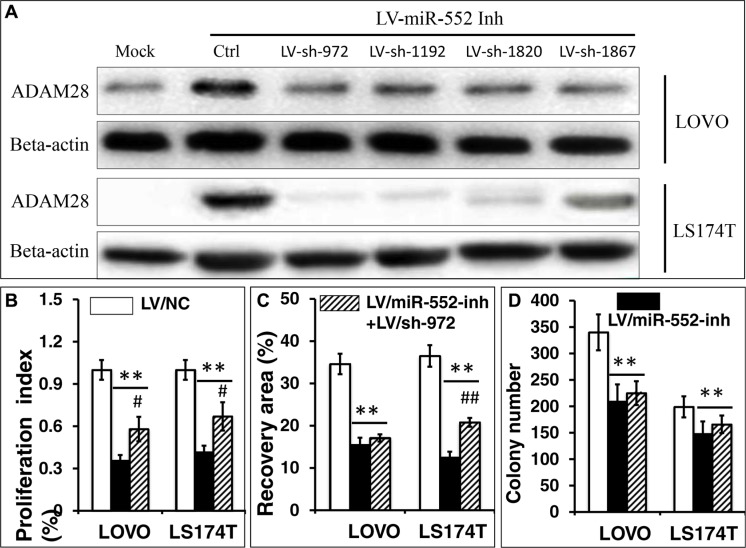
Colorectal cancer (CRC) is one of the most common prevalent cancer types worldwide. MicroRNAs (miRNAs or miRs) have been demonstrated to play crucial roles in the development, metastasis and drug resistance of CRC. In the present study, a strikingly elevated expression of miR-552 was determined in CRC tumor tissues and cells by a miRNA profiling analysis. Importantly, the gene of A Disintegrin And Metalloprotease (ADAM) family member 28 (ADAM28) was identified as a target of miR-552, which was further validated in terms of genetic dual luciferase report assay. Furthermore, an inhibition of miR-552 in LOVE and LS174T CRC cells by transducing miR-552 inhibitor (antagomiR-552) with a lentiviral vector exhibited an ability to reduce cell proliferation, migration and clonogenicity. Moreover, both LOVO and LS174T cells stably expressing miR-552 inhibitor displayed a decreased ability to develop tumors in a murine xenograft model in vivo. In contrast, a knockdown of ADAM28 by short hairpin RNA could reverse the antagomiR-552-induced inhibition of metastatic features of CRC cells in vitro. These results suggested that miR-552 is an oncomir able to promote CRC metastasis in part through a mechanism of targeting ADAM28, which may be a novel target for CRC treatment and warrants for further investigation.

Keywords: ADAM28; antagomir; colorectal cancer; miR-552; microRNAs.


Conflict of interest statement

CONFLICTS OF INTEREST

The authors declare that they have no competing interests.

Figures

Figure 1
Figure 1. Aberrant miR-552 and miR-592 transcripts in colorectal cancer tissues and cell lines
(A) Relative expression of miR-552 and miR-592 in CRC tissues. Both miR-552 and miR-592 expression is significantly elevated in CRC tissues as compared with the matched adjacent non-tumor tissues (p < 0.001). (B) Correlation of miR-552 and miR-592 in CRC tissues (r = 0.3568, 95% CI = 0.079–0.583, N = 50). (C) Relative expression of miR-552 and miR-592 in CRC cell lines. Both microRNAs were up-regulated in CRC cell lines HCT116, LOVO, LS174T and SW480, as compared with the CCD-18Co normal intestinal epithelial cell line (p < 0.001). Data expressed the mean ± SD; ***p < 0.001.
Figure 2
Figure 2. Immunohistochemistry (IHC) staining determined ADAM28 expression in human CRC tumors and matched adjacent tissues
(AF) Representative images of the expression of ADAM28 protein determined by an IHC staining. (A–C) Images represented the ADAM28 expression in non-CRC tumor adjacent tissues at different magnifications; (D–F) Images represented the ADAM28 expression in CRC tumor tissues at different magnifications. (G) Immunoblotting assay determined ADAM28 protein in 4 paired CRC tissues and non-tumor intestinal tissues (T = CRC tumor tissue; N = non-tumor intestinal tissues). (H) Semi-quantitative analysis of ADAM28 protein expression in the 4 paired tumor and non-tumor tissues in the Figure 4G by densitometry assay. Data represented ratio over respective loading control b-actin (P: Patient with CRC). (I) Relative expression of ADAM28 mRNA in paired CRC tumor tissues and the match non-tumor adjacent tumor tissues (N = 14). (J) Semi-quantitative analysis of ADAM28 protein expression using integrated absorbance (IA) in human CRC tissues and the matched adjacent non-tumor tissues. Value was expressed as the average values from each individual sample of CRC tumor tissues or its matched adjacent tissue. The total average value of IA in the CRC tumor tissues was significantly less abundant as compared with the matched adjacent tissues (p < 0.05, n = 14). Data was expressed as mean ± SD for 14 sets of samples. Bar in A, E: 100 μm; in B, C, F: 50 μm; D: 200 μm.
Figure 3
Figure 3. Validation of ADAM28 mRNA as a target of miR-552
(A) Sequence of potential binding site of miR-552 in the 3′UTR of human ADAM28 mRNA (top sequence), mutations were introduced into the binding site for generation of mutated ADAM28 3′TUR (bottom sequence). (B) Validation of miR-552 target using ADAM28 3′UTR luciferase reporter. C: The expression of ADAM28 was detected by immunoblotting in CCD-18Co normal colon epithelial cells transfected with indicated miR-552 control (NC), inhibitor and mimic. The immunoblotting assay showed an increased and decreased abundance of ADAM28 protein in the CCD-18Co cells transfected with the miR-552 inhibitor and mimic, respectively (top panel); in addition, an increased abundance of ADAM28 protein in the both LOVO and LS174T cells infected with the LV-miR-552 inh viral vector as compared to the LV-NC (top panel). **Compared with pSicoR/NC group, p < 0.01. Results represented the mean ± SD from three independent triplicated experiments (N = 9).
Figure 4
Figure 4. Generation of CRC cell lines expressing miR-552 inhibitor
Following a DNA cloning strategy, the proviral plasmids were used for production of lentiviral vector LV-miR-552-inh and LV-NC, which express miR-552 inhibitor and scramble control, respectively. These vectors also expressed an EGFP reporter gene for accessing the transduction efficiency. (AB) The LOVO cells transduced with LV-NC (A) and mR-552-inh (B) showed an infectivity of lentiviral vectors. (CD) The LS174T cells transduced with LV-NC (C) and mR-552-inh (D) showed an infectivity of lentiviral vectors. (E) qRT-PCR result exhibited a reduced abundance of miR-552 transcript in the LV-miR-552-inh-infected cells relative to the LV-NC-infected cells. Data represents the mean ± SD from three independent experiments. Compared to the LV-NC-infected group, ***p < 0.001.
Figure 5
Figure 5. Inhibition of miR-552 reduces LOVO and LS174T CRC cell migration and proliferation in vitro
LOVO and LS174T cells were infected with LV-miR-552-inh or LV-NC, the capability of cell migration was accessed in terms of a scratch assay, and cell proliferative ability was ascertained by an MTT method. (A) Representative images of scratch assays for LOVO cells (top two panels) and LS174T cells (bottom two panels) treated as indicated condition. (B) Relevant quantification of the results of cell migration index. (C) An MTT assay showed an inhibition of cell proliferation in LV-miR-552-inh-infected cells. Compared with LV-NC group, **p < 0.01. Data in B and C represented the mean ± SD from three independent triplicated experiments (N = 9).
Figure 6
Figure 6. Inhibition of miR-552 suppresses the clonogenicity in LOVO and LS174T CRC cells
LOVO and LS174T cells stably expressing miR-552-inh or miRNA NC were the formation of cell colonies using a clonogenic assay in 35-mm dishes. (A) Representative images of clonogenic assay for LOVO cells (top panel) and LS174T cells (bottom panel). (B) Relevant quantification of the results of cell clonogenic index, which showed an inhibition of clonogenic capacity in LV-miR-552-inh-infected cells. Compared with LV-NC group, **p < 0.01. Data in B represented the mean ± SD from three independent triplicated experiments (N = 9).
Figure 7
Figure 7. In vivo tumorigenic analysis of LV-miR-552-inh-infected CRC cells
SCID mice were subcutaneously injected 200 μl Matrigel containing 106 LV-infected LOVO or LS174T cells, and the formation of tumor was at three weeks after the injection. (A) Images showed sizes of tumors formed from LOVO cells infected with LV-miR-552-inh (top panel) and LV-NC (bottom panel). (B) Images showed sizes of tumors formed from LS174T cells infected with LV-miR-552-inh (top panel) and LV-NC (bottom panel). (C) Relevant quantification of the sizes (diameter) of tumors derived from CRC cells expressing miR-552-inh. The result showed an ability of miR-552 inhibitor to reduced tumor sizes. Compared with LV-NC group, **p < 0.01. Data in C represented the mean ± SD from 12 animals from two independent experiments (N = 12). Bars: 1.0 cm.
Figure 8
Figure 8. Knockdown of ADAM28 expression enhances the proliferation, migration and clonogenicity in LOVO and LS174T CRC cells
LV-miR-552-inh transduced LOVO and LS174T cells were co-infected with one of LV-shADAM28-972, LV-shADAM28-1192, LV-shADAM28-1820 or LV-shADAM28-1867, the expression of ADAM28 protein was determined by an immunoblotting assay (A), and the capacity of proliferation (B), cell migration (C) and clonogenicity (D) were accessed in terms of an MTT assay, a scratch assay, and clone formation assay, respectively. (A) Representative blots of AMAM28 expression in LOVO cells (top two panels) and LS174T cells (bottom two panels) following shRNA interfaces. The immunoblotting assay showed the LV-shADAM28-972 could efficiently knockdown the LV-miR-552-inh-restored ADAM28 in both LOVO and LS174T cells, which was chosen for using in further analysis in this study. (B) An MTT assay showed a promotion of cell proliferation in LV-shADAM28-972 and LV-miR-552-inh-co-infected LOVO and LS174T cells (p < 0.05). (C) Relevant quantification of the results of cell migration index showed a significantly enhanced capacity of migration in LV-shADAM28-972 and LV-miR-552-inh-co-infected LS174T cells (p < 0.05), but not LOVO cells. (D) Relevant quantification of the results of cell clonogenic index, which showed a moderate increase of clonogenic capacity in LV-shADAM28-972 and LV-miR-552-inh-co-infected cells (p > 0.05). Compared with LV-NC group, *p < 0.01; **p < 0.01. Compared with LV-miR-552-inh, #p < 0.01; ##p < 0.01. Data in B–D represented the mean ± SD from three independent triplicated experiments (N = 9).

References

    1. Siegel RL, Miller KD, Jemal A. Cancer statistics, 2015. CA Cancer J Clin. 2015;65:5–29. - PubMed
    1. Garofalo M, Croce CM. MicroRNAs as therapeutic targets in chemoresistance. Drug resistance updates. 2013;16:47–59. - PMC - PubMed
    1. Cojoc M, Mabert K, Muders MH, Dubrovska A. A role for cancer stem cells in therapy resistance: cellular and molecular mechanisms. Seminars in cancer biology. 2015;31:16–27. - PubMed
    1. Kartal-Yandim M, Adan-Gokbulut A, Baran Y. Molecular mechanisms of drug resistance and its reversal in cancer. Critical reviews in biotechnology. 2015:1–11. - PubMed
    1. Lippert TH, Ruoff HJ, Volm M. Intrinsic and acquired drug resistance in malignant tumors. The main reason for therapeutic failure. Arzneimittel-Forschung. 2008;58:261–264. - PubMed