. 2016 Sep 23;11(9):e0163332.
doi: 10.1371/journal.pone.0163332. eCollection 2016.

Feasibility of MR-Based Body Composition Analysis in Large Scale Population Studies

Affiliations

Feasibility of MR-Based Body Composition Analysis in Large Scale Population Studies

Janne West et al. PLoS One. .

Abstract

Introduction: Quantitative and accurate measurements of fat and muscle in the body are important for prevention and diagnosis of diseases related to obesity and muscle degeneration. Manually segmenting muscle and fat compartments in MR body-images is laborious and time-consuming, hindering implementation in large cohorts. In the present study, the feasibility and success-rate of a Dixon-based MR scan followed by an intensity-normalised, non-rigid, multi-atlas based segmentation was investigated in a cohort of 3,000 subjects.

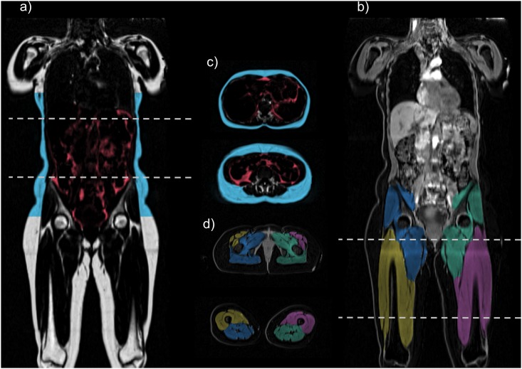
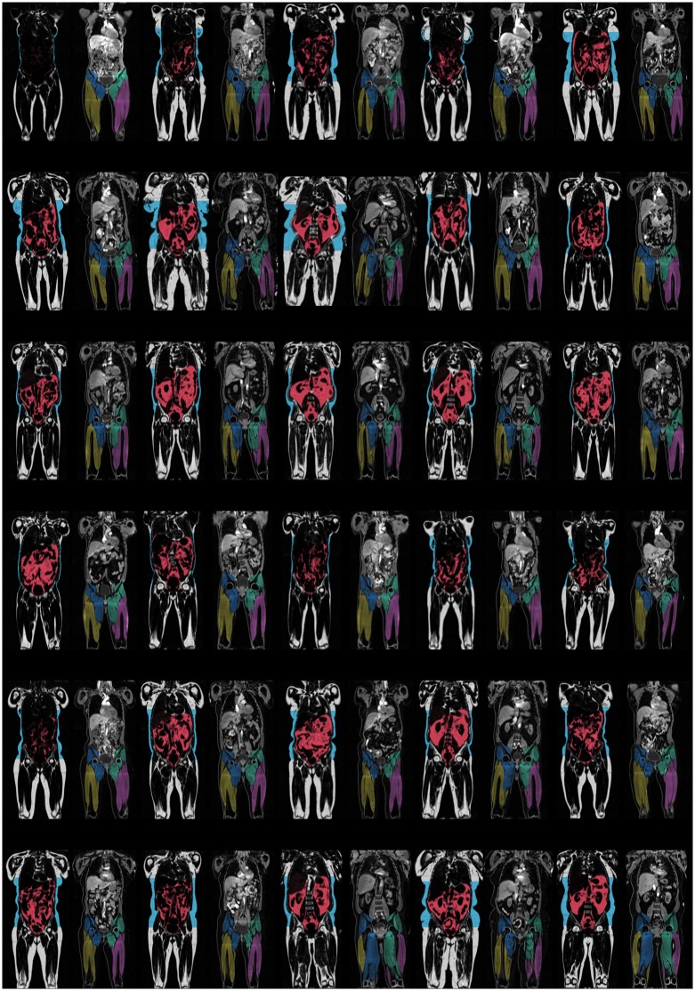
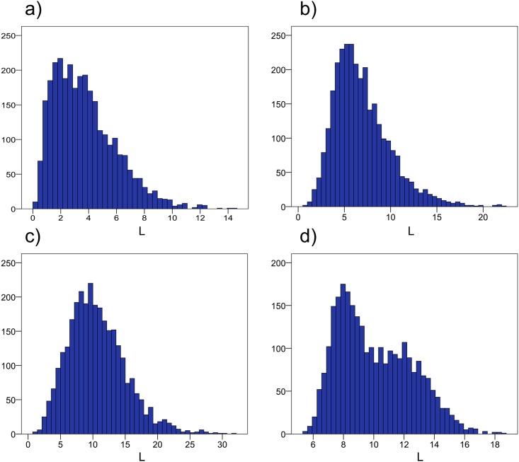
Materials and methods: 3,000 participants in the in-depth phenotyping arm of the UK Biobank imaging study underwent a comprehensive MR examination. All subjects were scanned using a 1.5 T MR-scanner with the dual-echo Dixon Vibe protocol, covering neck to knees. Subjects were scanned with six slabs in supine position, without localizer. Automated body composition analysis was performed using the AMRA Profiler™ system, to segment and quantify visceral adipose tissue (VAT), abdominal subcutaneous adipose tissue (ASAT) and thigh muscles. Technical quality assurance was performed and a standard set of acceptance/rejection criteria was established. Descriptive statistics were calculated for all volume measurements and quality assurance metrics.

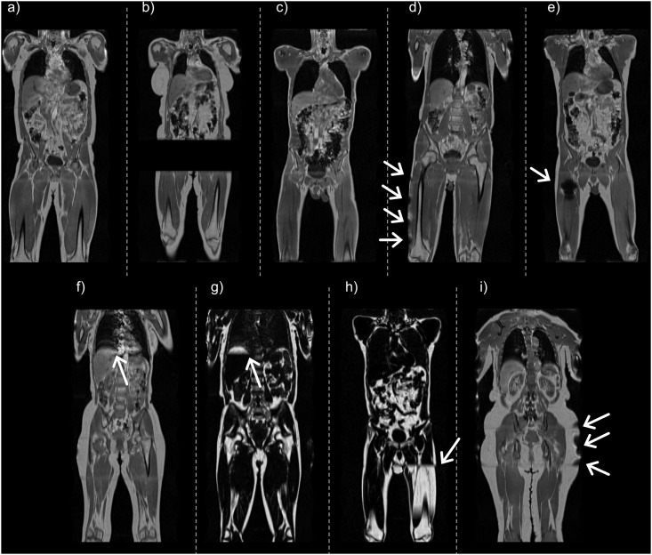
Results: Of the 3,000 subjects, 2,995 (99.83%) were analysable for body fat, 2,828 (94.27%) were analysable when body fat and one thigh was included, and 2,775 (92.50%) were fully analysable for body fat and both thigh muscles. Reasons for not being able to analyse datasets were mainly due to missing slabs in the acquisition, or patient positioned so that large parts of the volume was outside of the field-of-view.

Discussion and conclusions: In conclusion, this study showed that the rapid UK Biobank MR-protocol was well tolerated by most subjects and sufficiently robust to achieve very high success-rate for body composition analysis. This research has been conducted using the UK Biobank Resource.


Conflict of interest statement

JW, ODL, TR, and MB are stock holders in Advanced MR Analytics (AMRA). This does not alter our adherence to PLOS ONE policies on sharing data and materials.

Figures

Fig 1
Fig 1. Typical imaging artifacts within the UK Biobank imaging study.
(a) tall subject where the complete femoral epicondyles are not visible, thighs are not analysable, (b) missing slab over the abdominal and thigh regions, not analysable, (c) misplaced landmark where the complete femoral epicondyles are not within the imaged volume, thighs are not analysable, (d) tilted subject where the right thigh is partly outside the field-of-view, right thigh not analysable (e) metal artifact, right thigh not analysable, (f) respiratory artifact, visible as ghosting on top of liver, (g) swap in top-of-liver, (h) separate-island swap, this was corrected, and (i) outer field-of-view inhomogeneities. Characteristic artifacts are highlighted with arrows.
Fig 2
Fig 2. Coronal and transverse slices of one subject.
(a) intensity-corrected coronal fat image with fat segmentations using overlay colours, (b) intensity-corrected coronal water image with muscle segmentations using overlay colours, (c) corresponding transverse slices with fat segmentation, and (d) corresponding transverse slices with muscle segmentation.
Fig 3
Fig 3. Central coronal slices from 1% of the imaged subjects.
Demonstrating the wide range of phenotypes within the UK Biobank imaging study. For each subject; left shows intensity-corrected coronal fat image with fat segmentations using overlay colours, right shows intensity-corrected coronal water image with muscle segmentations using overlay colours.
Fig 4
Fig 4. Histograms from all 3,000 subjects.
(a) visceral adipose tissue (VAT), (b) abdominal subcutaneous tissue (ASAT), (c) total trunk fat (defined as VAT+ASAT), and (d) total thigh volumes.

References

    1. Urbina EM, Kimball TR, McCoy CE, Khoury PR, Daniels SR, Dolan LM. Youth with obesity and obesity-related type 2 diabetes mellitus demonstrate abnormalities in carotid structure and function. Circulation. 2009;119(22):2913–9. 10.1161/CIRCULATIONAHA.108.830380 - DOI - PMC - PubMed
    1. WHO. Global status report on noncommunicable diseases 2014. Geneva, Switzerland: WHO Press; 2014. 298 p.
    1. Artham SM, Lavie CJ, Patel HM, Ventura HO. Impact of obesity on the risk of heart failure and its prognosis. Journal of the cardiometabolic syndrome. 2008;3(3):155–61. 10.1111/j.1559-4572.2008.00001.x . - DOI - PubMed
    1. Ninomiya JK, L'Italien G, Criqui MH, Whyte JL, Gamst A, Chen RS. Association of the metabolic syndrome with history of myocardial infarction and stroke in the Third National Health and Nutrition Examination Survey. Circulation. 2004;109(1):42–6. 10.1161/01.CIR.0000108926.04022.0C . - DOI - PubMed
    1. Neeland IJ, Ayers CR, Rohatgi AK, Turer AT, Berry JD, Das SR, et al. Associations of visceral and abdominal subcutaneous adipose tissue with markers of cardiac and metabolic risk in obese adults. Obesity. 2013;21(9):E439–47. 10.1002/oby.20135 - DOI - PMC - PubMed