. 2016 Nov 1;23(11):1815-1826.
doi: 10.1038/cdd.2016.101. Epub 2016 Sep 23.

Leishmania donovani inhibits macrophage apoptosis and pro-inflammatory response through AKT-mediated regulation of β-catenin and FOXO-1

Affiliations

Leishmania donovani inhibits macrophage apoptosis and pro-inflammatory response through AKT-mediated regulation of β-catenin and FOXO-1

Purnima Gupta et al. Cell Death Differ. .

Abstract

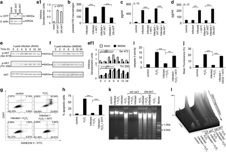
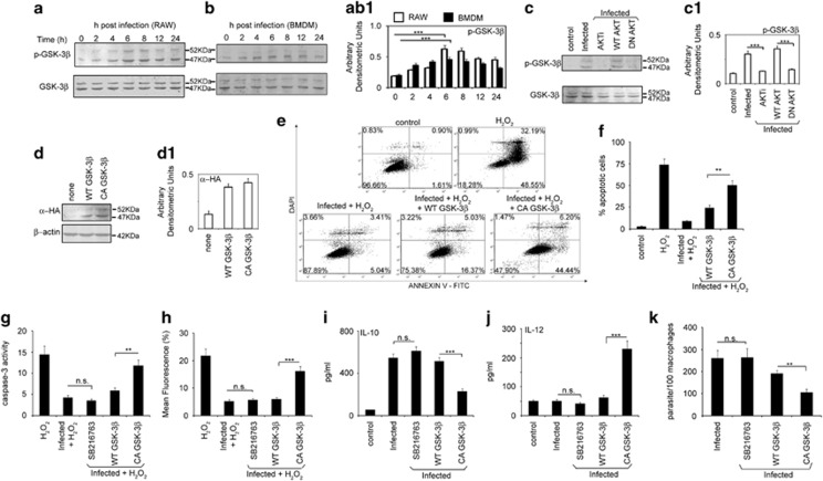
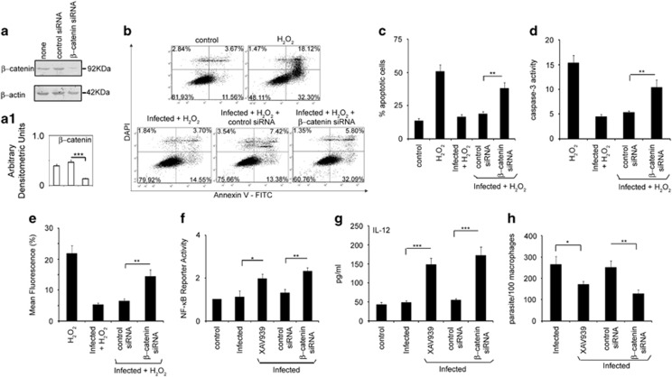
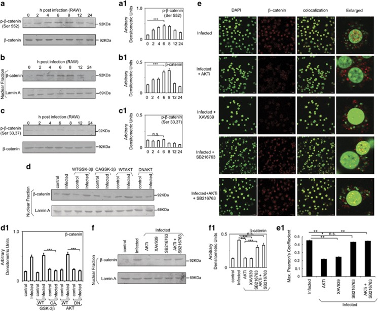
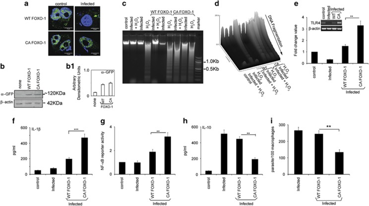
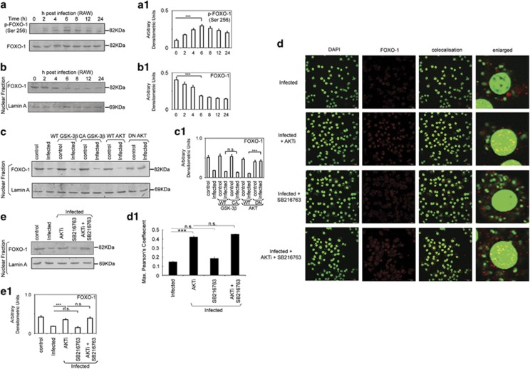
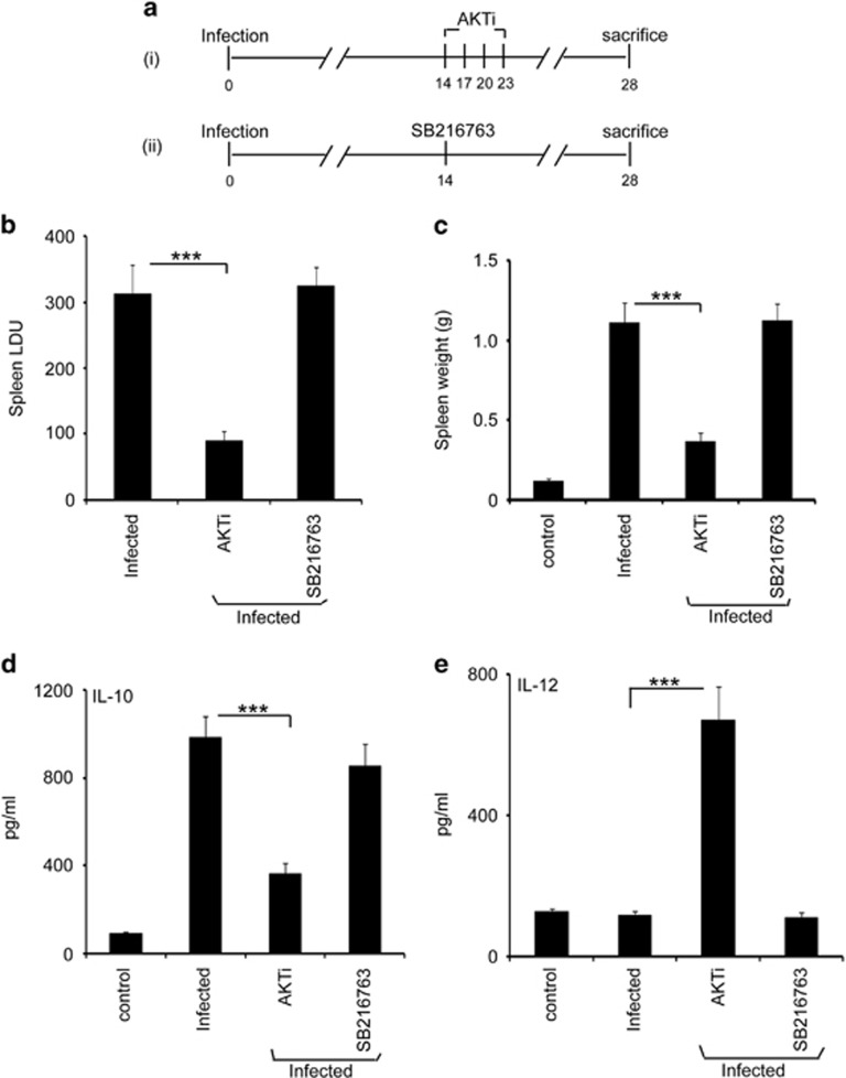
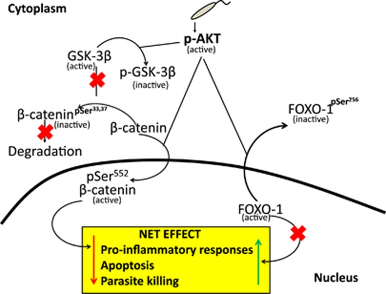
In order to establish infection, intra-macrophage parasite Leishmania donovani needs to inhibit host defense parameters like inflammatory cytokine production and apoptosis. In the present study, we demonstrate that the parasite achieves both by exploiting a single host regulator AKT for modulating its downstream transcription factors, β-catenin and FOXO-1. L. donovani-infected RAW264.7 and bone marrow-derived macrophages (BMDM) treated with AKT inhibitor or dominant negative AKT constructs showed decreased anti-inflammatory cytokine production and increased host cell apoptosis resulting in reduced parasite survival. Infection-induced activated AKT triggered phosphorylation-mediated deactivation of its downstream target, GSK-3β. Inactivated GSK-3β, in turn, could no longer sequester cytosolic β-catenin, an anti-apoptotic transcriptional regulator, as evidenced from its nuclear translocation during infection. Constitutively active GSK-3β-transfected L. donovani-infected cells mimicked the effects of AKT inhibition and siRNA-mediated silencing of β-catenin led to disruption of mitochondrial potential along with increased caspase-3 activity and IL-12 production leading to decreased parasite survival. In addition to activating anti-apoptotic β-catenin, phospho-AKT inhibits activation of FOXO-1, a pro-apoptotic transcriptional regulator. Nuclear retention of FOXO-1, inhibited during infection, was reversed when infected cells were transfected with dominant negative AKT constructs. Overexpression of FOXO-1 in infected macrophages not only documented increased apoptosis but promoted enhanced TLR4 expression and NF-κB activity along with an increase in IL-1β and decrease in IL-10 secretion. In vivo administration of AKT inhibitor significantly decreased liver and spleen parasite burden and switched cytokine balance in favor of host. In contrast, GSK-3β inhibitor did not result in any significant change in infectivity parameters. Collectively our findings revealed that L. donovani triggered AKT activation to regulate GSK-3β/β-catenin/FOXO-1 axis, thus ensuring inhibition of both host cell apoptosis and immune response essential for its intra-macrophage survival.


Figures

Figure 1
Figure 1
Role of AKT in L. donovani infection. (ad) RAW264.7 cells (2 × 10⁶) were treated with either AKTi (10 μM) for 1 h or transiently transfected with WT- or DN-AKT expression plasmids for 24 h. Both these macrophages were then infected with L. donovani promastigotes (macrophage:parasite ratio, 1:10) for 24 h. Expression of GFP in transfected cells were detected by Western blotting (a) and intracellular parasite number (b) were determined by DAPI staining whereas levels of IL-10 (c) and IL-12 (d) were measured by ELISA. (e and f) Both RAW264.7 (e) and BMDM (f) were infected with L. donovani promastigotes for different time periods as indicated. Levels of phosphorylated and total AKT were then detected by Western Blotting. (gj) RAW cells were treated with AKTi for 1 h, infected with L. donovani promastigotes for 6 h and then treated with 400 μM H2O2 for 1 h. These cells were analyzed for the extent of apoptosis by annexin V-tagged FITC-DAPI flow cytometry after washing and incubation for 24 h at 37 °C (g and h). Dual parameter dot plot of FITC fluorescence (x axis) versus DAPI fluorescence (y axis) is represented as logarithmic fluorescence intensity. Quadrants are as follows: upper left, necrotic cells; lower left, live cells; lower right, apoptotic cells; upper right, necrotic or late phase of apoptotic cells. After washing, whole cell lysate of these cells (10 μg of protein per sample) were used to determine caspase-3 activity using Ac-DEVDpNA as substrate (i). Mitochondrial integrity was measured in these AKTi treated infected-H2O2 treated cells after incubation with DiOC6 (40 nM) for an additional 30 min (j). (k) RAW264.7 cells were transiently transfected with either WT- or DN-AKT expression plasmids, infected with L. donovani promastigotes for 6 h and then treated with H2O2 (400 μM) for 1 h. DNA fragmentation profile was analyzed by agarose gel electrophoresis.(l) A representative surface plot analyzed by using ImageJ software indicates the extent of fragmentation. All experiments were repeated at least three times each and one set of representative data is shown. Bands were analyzed densitometrically and bar graphs expressing arbitrary densitometric units are presented adjacent to corresponding western blots. Error bars represent mean±S.D., n=3.**P<0.01, ***P<0.001; Student's t-test
Figure 2
Figure 2
Role of GSK-3β in L. donovani infection. (a and b) Both RAW264.7 (a) and BMDM (b) were infected with L. donovani promastigotes (macrophage:parasite ratio, 1:10) for different time periods as indicated. Levels of phosphorylated and total GSK-3β were then detected by Western Blotting. (c) Cell lysates were prepared from AKTi treated or WT- or DN-AKT transfected infected cells and levels of phosphorylated and total GSK-3β were detected by immunoblotting. (dh) RAW264.7 cells were either treated with SB216763 (20 μM) for 1 h or transfected with WT- or CA-GSK-3β expression plasmids, infected with L. donovani promastigotes for 6 h and then treated with 400 μM H2O2 for 1 h. Expression of Hemagglutinin (HA) in whole cell lysates were analyzed in transfected cells by Western blotting (d). Similarly, transfected cells were analyzed for the extent of apoptosis by annexin V-tagged FITC-DAPI flow cytometry (e and f), caspase-3 activity using Ac-DEVDpNA as substrate (g) and mitochondrial integrity after incubation with DiOC6 (40 nM) (h) as described in the legend of Figure 1. (i–k) Macrophages were treated with SB216763 or transfected with WT-or CA-GSK-3β expression plasmids, infected with L. donovani promastigotes for 24 h and processed for determination of either IL-10 (i) and IL-12 (j) by ELISA or checked for parasite survival (k). All experiments were repeated at least three times each and one set of representative data is shown. Bands were analyzed densitometrically and bar graphs expressing arbitrary densitometric units are presented adjacent to corresponding western blots. Error bars represent mean±S.D., n=3. NS, non-significant, **P<0.01, ***P<0.001; Student's t-test
Figure 3
Figure 3
Role of β-catenin in apoptois and inflammatory responses during infection. (a) RAW264.7 cells were transfected (24 h) with either control or β-catenin siRNA followed by infection with L. donovani promastigotes for 6 h. Expression of β-catenin was evaluated by immunoblot analysis. (b-e) Macrophages were transfected with β-catenin siRNA for 24 h, infected with L. donovani promastigotes for 6 h and then treated with 400 μM H2O2 for 1 h. These cells were then analyzed for the extent of apoptosis by annexin V-tagged FITC-DAPI flow cytometry after incubation for 24 h at 37 °C (b and c), caspase-3 activity using Ac-DEVDpNA as substrate (d) and mitochondrial integrity (e) as described in the legend of Figure 1. (f) RAW264.7 cells were treated with XAV939 (1 μM) or transfected with control or β-catenin siRNA along with pNF-κB luciferase plasmid (1 μg) and 0.5 μg of pCMV-β-gal. After 24 h of transfection, cells were infected with L. donovani promastigotes for 24 h, washed, lysed and processed for luciferase activity. (g and h) XAV939-treated or β-catenin siRNA transfected cells were infected with L. donovani promastigotes for 24 h and were processed for determination of IL-12 by ELISA (g) or estimation of extent of parasite survival (h). All experiments were repeated at least three times each and one set of representative data is shown. Bands were analyzed densitometrically and bar graphs expressing arbitrary densitometric units are presented adjacent to corresponding western blots. Error bars represent mean±S.D., n=3. *P<0.05, **P<0.01, ***P<0.001; Student's t-test
Figure 4
Figure 4
Role of AKT and GSK-3β in modulation of β-catenin localization in infection. (a) RAW264.7 cells were infected with L. donovani promastigotes (macrophage:parasite ratio, 1:10) for different time periods as indicated. Levels of phosphorylated and total β-catenin were then detected by Western Blotting using respective antibodies. (b) Nuclear fractions were isolated from RAW264.7 cells infected with L. donovani promastigotes for different times and β-catenin expression was checked by immunoblotting. (c) Infected RAW264.7 cells were lysed after different time periods and checked for the levels of phosphorylated and total β-catenin by Western Blotting using respective antibodies. (d) RAW264.7 cells were transfected (24 h) with WT-AKT, DN-AKT, WT-GSK-3β and CA-GSK-3β expression plasmids followed by infection with L. donovani promastigotes for 6 h. Nuclear fractions were isolated and expression of β-catenin was evaluated by immunoblot analysis. (e) Macrophages were treated with AKTi or SB216763 or XAV939 for 1 h followed by infection with L. donovani promastigotes for 6 h. Cells were then stained with anti-β-catenin monoclonal antibody followed by secondary Alexa555-conjugated antibody. Nuclei were stained with DAPI and cells were analyzed under microscope. Images were analyzed for colocalization using Olympus Fluoview (version 3.1a; Tokyo, Japan). For better visual aid green was used instead of blue to mark DAPI using LUT program of Fluoview. Red or yellow colors on green nucleus denotes strong colocalization, whereas green nucleus lacking any red or yellow colors denotes exclusion. (E1) Maximum Pearson's Coeffiecient was calculated by randomly selecting at least 15 cells per field for at least three random fields per experiment using colocalization program of Olympus Fluoview (version 3.1a; Tokyo, Japan). (f) Macrophages were treated with AKTi and/or SB216763 or XAV939 and infected for 6 h. Thereafter nuclear fractions were isolated and expression of β-catenin was assessed. Lamin serves as control for the nuclear extract. All experiments were repeated at least three times each and one set of representative data is shown. Bands were analyzed densitometrically and bar graphs expressing arbitrary densitometric units are presented adjacent to corresponding western blots. Error bars represent mean±S.D., n=3. NS, non-significant, **P<0.01,***P<0.001; Student's t-test
Figure 5
Figure 5
Role of FOXO-1 in apoptosis and inflammatory response during infection. (a) RAW264.7 cells were transfected with either GFP-WT-FOXO-1 or GFP-CA-FOXO-1 and infected with L. donovani promastigotes for 6 h. Cells were fixed and stained with DAPI and analyzed under microscope. (bd) Cells were transiently transfected with WT- or CA-FOXO-1 expression plasmid (24 h) and then infected with L. donovani for 6 h. Transfection efficiency was further analyzed by checking the expression of GFP in whole cell lysates (b). Transfected and L. donovani-infected cells were treated with H2O2 for 1 h, washed and incubated for additional 24 h and the integrity of DNA was analyzed with the appearance of DNA ladders in each group via agarose electrophoresis (c). Representative surface plot indicates the extent of fragmentation (d). (ei) Cells were transfected with the indicated constructs and infected with parasites for 24 h. This was followed by real-time PCR analysis of TLR4 levels as fold change (e) or processed for determination of IL-1β (f) and IL-10 (g) levels by ELISA. RAW264.7 cells were transfected with desired constructs along with pNF-κB luciferase plasmid (1 μg) and 0.5 μg of pCMV-β-gal. After 24 h of transfection, cells were infected with L. donovani for 24 h, lysed, and processed for luciferase activity (h). The parasite survival was determined by staining the cells with DAPI after transfection with indicated constructs and counting parasites per 100 macrophages (i). All experiments were repeated at least three times each and one set of representative data is shown. Bands were analyzed densitometrically and bar graphs expressing arbitrary densitometric units are presented adjacent to corresponding western blots. Error bars represent mean±S.D., n=3. **P<0.01, ***P<0.001; Student's t-test
Figure 6
Figure 6
Role of AKT and GSK-3β in modulation of FOXO-1 localization. (a and b) RAW264.7 cells were infected for different times and phospho-FOXO-1 expression at protein level was checked by immunoblotting in whole cell lysate (a) whereas FOXO-1 level was checked in nuclear fractions (b). (c) RAW cells were transfected with WT-GSK-3β or CA-GSK-3β or WT-AKT or DN-AKT for 24 h and then infected with L. donovani promastigotes for 6 h. Cells were lysed, nuclear fractions were obtained and FOXO-1 expression at protein level was checked by immunoblotting. (d) Cells were treated with AKTi or SB216763 for 1 h followed by infection with L. donovani promastigotes for 6 h. Cells were stained with anti-FOXO-1 monoclonal antibody followed by Alexa555-conjugated secondary antibody. Nuclei were stained with DAPI, and cells were analyzed as mentioned in legends of Figure 4. (e) Macrophages were treated with AKTi and/or SB216763 and infected for 6 h. Thereafter nuclear fractions were isolated and expression of FOXO-1 was assessed. Lamin serves as control for the nuclear extract. All experiments were repeated at least three times each and one set of representative data is shown. Bands were analyzed densitometrically and bar graphs expressing arbitrary densitometric units are presented adjacent to corresponding western blots. Error bars represent mean±S.D., n=3. NS, non-significant, ***P<0.001; Student's t-test
Figure 7
Figure 7
In vivo validation of the role of AKT in infection. (a) Schematic representation of the experimental protocol for treatment of L. donovani-infected mice: (i) infected and AKTi treated and (ii) infected and SB216763-treated. (b and d) Infected mice were treated as mentioned in the scheme (a) and disease progression was determined by measuring spleen parasite burden as Leishman–Donovan units (b). Spleen was isolated and spleen weight was measured (c). (d and e) Splenocytes were isolated from control, infected, and infected plus AKTi-treated or SB216763-treated mice at 4 wk post-infection and IL-10 (d) and IL-12 (e) levels were determined by ELISA. Animal experiments were done with five animals per group. Error bars represent mean±S.D., n=3. ***P<0.001; Student's t-test
Figure 8
Figure 8
Modulation of host AKT pathway by L. donovani for successful survival. L. donovani activates host AKT by phosphorylation on serine and threonine residues, which in turn imparts a dual effect on the downstream signaling pathway: 1. Inactivation of GSK-3β by phosphorylation at Ser9 residue resulting in inhibition of Ser33/37 phosphorylation-mediated degradation of β-catenin and 2. Phosphorylation of β-catenin at Ser552 leading to its nuclear localization with concomitant inactivation of FOXO-1 by phosphorylation at Ser256 causing its cytosolic translocation. Overall these effects help parasite survival by inhibiting host cell apoptosis on one hand and modulating inflammatory responses on the other

Similar articles

Cited by

References

    1. Bhardwaj S, Srivastava N, Sudan R, Saha B. Leishmania interferes with host cell signaling to devise a survival strategy. J Biomed Biotechnol 2010; 2010: 109189. - PMC - PubMed
    1. Cecilio P, Pérez-Cabezas B, Santarém N, Maciel J, Rodrigues V, Cordeiro da Silva A et al. Deception and manipulation: the arms of Leishmania, a successful parasite. Front Immunol 2014; 5: 480. - PMC - PubMed
    1. Zhang Y, Wang X, Yang H, Liu H, Lu Y, Han L et al. Kinase AKT controls innate immune cell development and function. Immunology 2013; 140: 143–152. - PMC - PubMed
    1. Neves BM, Silvestre R, Resende M, Ouaissi A, Cunha J, Tavares J et al. Activation of phosphatidylinositol 3-kinase/Akt and impairment of nuclear factor-kappaB: molecular mechanisms behind the arrested maturation/activation state of Leishmania infantum-infected dendritic cells. Am J Pathol 2010; 177: 2898–2911. - PMC - PubMed
    1. Ruhland A, Leal N, Kima PE. Leishmania promastigotes activate PI3K/Akt signalling to confer host cell resistance to apoptosis. Cell Microbiol 2007; 9: 84–96. - PubMed

Publication types

MeSH terms