

Mycoplasma agalactiae Induces Cytopathic Effects in Infected Cells Cultured In Vitro
- PMID: 27662492
- PMCID: PMC5035028
- DOI: 10.1371/journal.pone.0163603
Mycoplasma agalactiae Induces Cytopathic Effects in Infected Cells Cultured In Vitro
Abstract
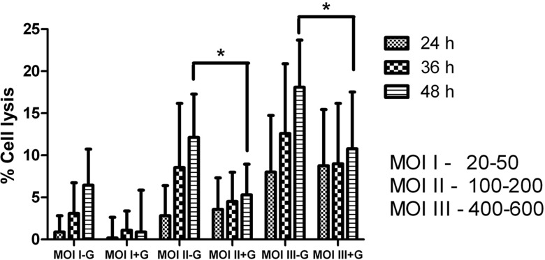
Mycoplasma agalactiae is the etiological agent of the contagious agalactia syndrome in sheep and goats and causes significant economic losses worldwide. Yet the mechanism of pathogenesis is largely unknown. Even whole-genome sequence analysis of its pathogenic type strain did not lead to any conclusions regarding its virulence or pathogenicity factors. Although inflammation and tissue destruction at the local site of M. agalactiae infection are largely considered as effects of the host immune response, the direct effect of the agent on host cells is not completely understood. The aim of this study was to investigate the effect of M. agalactiae infection on the quality and viability of host cells in vitro. Changes in cell morphology including cell elongation, cytoplasm shrinkage and membrane blebbing were observed in infected HeLa cells. Chromatin condensation and increased caspase-3 cleavage in infected HeLa cells 48 h after infection suggests an apoptosis-like phenomenon in M. agalactiae-infected cells. In compliance with these results, decreased viability and cell lysis of M. agalactiae-infected HeLa cells was also observed. Measurement of the amount of LDH released after M. agalactiae infection revealed a time- and dose-dependent increase in HeLa cell lysis. A significant decrease in LDH released after gentamicin treatment of infected cells confirmed the major role of cytadherent M. agalactiae in inducing host cell lysis. This is the first study illustrating M. agalactiae's induction of cytopathic effects in infected HeLa cells. Further detailed investigation of infected host tissue for apoptotic markers might demonstrate the association between M. agalactiae-induced host cell lysis and the tissue destruction observed during M. agalactiae natural infection.
Conflict of interest statement
The authors have declared that no competing interests exist.
Figures


References
-
- Rosengarten R, Citti C, Glew M, Lischewski A, Droesse M, Much P, et al. Host-pathogen interactions in mycoplasma pathogenesis: virulence and survival strategies of minimalist prokaryotes. Int J Med Microbil. 2000; 290(1): 15–25. - PubMed
-
- Rottem S. Interaction of mycoplasmas with host cells. Physiol Rev. 2003; 83(2): 417–432. - PubMed
MeSH terms
Grants and funding
LinkOut - more resources
Full Text Sources
Other Literature Sources
Research Materials
