. 2016 Nov 8;7(45):72923-72940.
doi: 10.18632/oncotarget.12141.

Grp94 in complexes with IgG is a soluble diagnostic marker of gastrointestinal tumors and displays immune-stimulating activity on peripheral blood immune cells

Affiliations

Grp94 in complexes with IgG is a soluble diagnostic marker of gastrointestinal tumors and displays immune-stimulating activity on peripheral blood immune cells

Elisa Tramentozzi et al. Oncotarget. .

Abstract

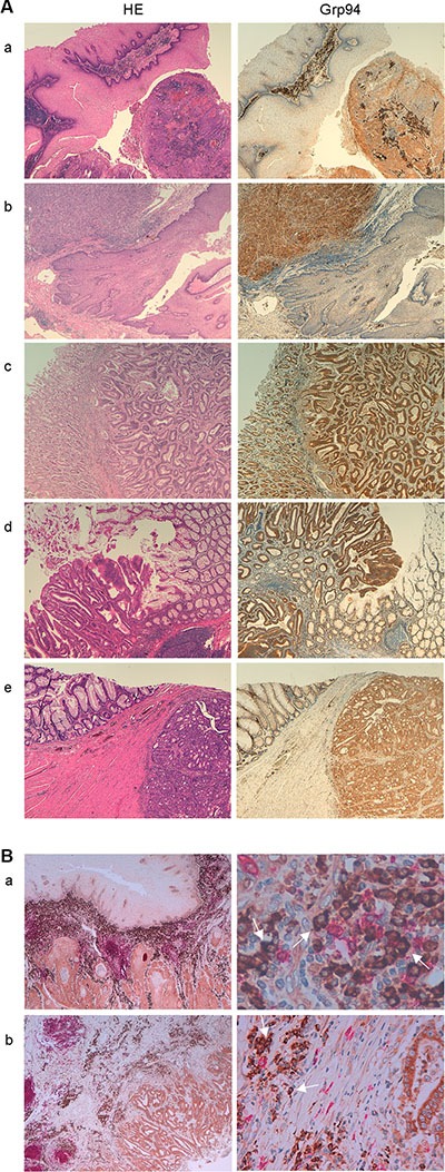
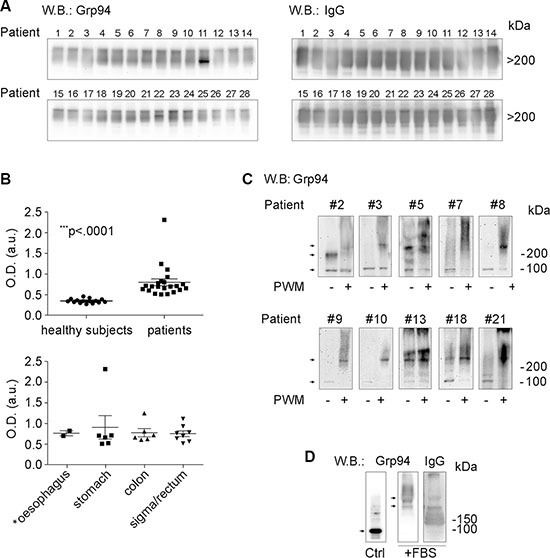
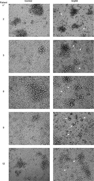
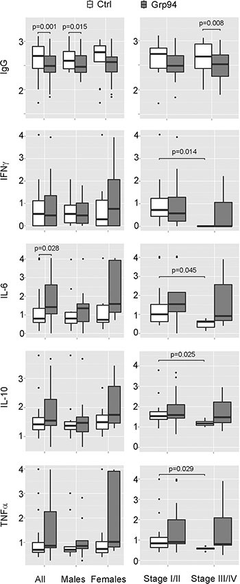
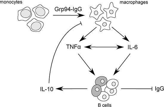
Glucose-regulated protein94 (Grp94), the most represented endoplasmic reticulum (ER)-resident heat shock protein (HSP), is a tumor antigen shared by different types of solid and hematological tumors. The tumor-specific feature of Grp94 is its translocation from the ER to the cell surface where it displays pro-oncogenic functions. This un-physiological location has important implications for both the tumor pathology and anti-tumor therapy. We wanted to address the question of whether Grp94 could be measured as liquid marker in cancer patients in order to make predictions of diagnostic and therapeutic relevance for the tumor. To this aim, we performed an in-depth investigation on patients with primary tumors of the gastrointestinal (GI) tract, using different methodological approaches to detect Grp94 in tumor tissues, plasma and peripheral blood mononuclear cells (PBMCs). Results indicate that Grp94 is not only the antigen highly expressed in any tumor tissue and in cells of tumor infiltrates, mostly B lymphocytes, but it is also found in the circulation. However, the only form in which Grp94 was detected in the plasma of any patients and in B lymphocytes induced to proliferate, was that of stable complexes with Immunoglobulin (Ig)G. Using a specific immune-enzyme assay to measure plasma Grp94-IgG complexes, we showed that Grp94-IgG complexes were significantly increased in cancer patients compared to healthy control subjects, serving as diagnostic tumor biomarker. Results also demonstrate that the stimulation of patient PBMCs with Grp94-IgG complexes led to an increased secretion of inflammatory cytokines that might drive a potentially beneficial anti-tumor effect.

Keywords: biomarkers; gastrointestinal neoplasms; heat shock Proteins; immunoglobulins; immunomodulation.


Conflict of interest statement

CONFLICTS OF INTEREST

The authors declare that they have no conflicts of interests.

Figures

Figure 1
Figure 1. Grp94 marks cancer cells of any tumor of the GI tract and cells of tumor infiltrates
(A) Specimens of different tumors (a, oesophageal squamous cell carcinoma; b, adeno-carcinoma of the gastro-enteric junction; c, tubular-type adeno-carcinoma of the stomach; d and e, large bowel adeno-carcinomas) were stained with H&E and incubated with rat monoclonal anti-Grp94 Abs for specific immunostaining, as specified in Methods. Magnifications are 10× (a–d) and 20× (e). (B) Double immunostaining for both CD20+ cells (anti-human CD20cy Abs) and Grp94 of specimens of: a, oesophageal squamous cell carcinoma (20×, left) showing diffuse infiltration of B cells into the tumor stroma with the enlargement (63×, right) showing that the Grp94-positive cells are mostly plasma cells (arrows); b, large bowel carcinoma (10×, left) with enlargement (40×, right) revealing the same features as in a.
Figure 2
Figure 2. Grp94 in complexes with IgG is the form in which the tumor antigen Grp94 circulates in plasma and is presented by plasma cells
(A) WB for Grp94 and IgG in plasma of all patients. Patients are numbered following the order by which they were analyzed. Ten μg of proteins of each plasma sample was loaded on to 4–20% polyacrylamide gel in denaturing conditions of PAGE, without boiling and reducing treatment of samples. WB was performed as specified in Methods, probing the membrane with primary anti-Grp94 monoclonal Abs (clone 9G10, Santa Cruz Biotech., Santa Cruz, CA, USA) followed by anti-rat HRP-conjugated secondary Abs, and with primary anti-human IgG polyclonal Abs followed by anti-sheep HRP-conjugated secondary Abs. Cropped WB of samples 1–14 and samples 15–28 originate from two distinct membranes. Patient #16 was then excluded from the following analyses for having a tumor that secondarily involved the large bowel. Only the Grp94-positive bands in the membranes are shown since no other band was visualized. Grp94 at masses higher than 200 kDa indicates the formation of complexes with IgG, as proved by the intense co-positivity for human IgG of the Grp94-positve bands (see WB on right). Specificity of the immune reaction was also evaluated by probing the membranes with only secondary Abs. (B) In the upper panel, scatter plots are shown of ELISA determinations of Grp94-IgG complexes in plasma of 15 healthy subjects and 22 cancer patients (13 males, 9 females). Measurements were made by testing each plasma sample in duplicate at the dilution of 1:128, and absorbance was measured at 450 nm (OD, optical density). Horizontal lines indicate the mean value with SE. Minimum and maximum OD values were, respectively, 0.27 and 0.458 in healthy subjects, and 0.506 and 2.31 in patients. The mean ± SE of OD values in healthy subjects was 0.346 ± 0.013, and in patients 0.796 ± 0.0817. Lower and upper 95% CI of the mean was 0.318 and 0.375 in healthy subjects, 0.627 and 0.966 in patients. The difference between the two groups was statistically significant (Mann-Whitney test, two-tailed). The lower panel shows the ELISA measurements in the same patients grouped by type of tumor. The mean ± SE OD values did not differ significantly among groups. The patient with the highest value of OD (2.31) died after two months from the admission. (C) Cropped WB for Grp94 on lysates of PBMCs of a representative number of patients (eight with large bowel adenocarcinomas and two with gastric carcinomas). Patients #3, #7, #9, #10 and #13 had tumors at stage III/IV. PBMCs were incubated in absence (control) and presence of the B cell stimulatory agent Pokeweed mitogen (PWM) at 20 μg/ml (final concentration), and after 10 days of incubation cells were lysed and treated as specified in Methods. The protein concentration was previously determined by means of calibration SDS-PAGE (10% polyacrylamide gel) and the same protein quantity (without boiling and reducing treatments) was loaded for each sample on to a 4–20% polyacrylamide gel and then probed with anti-Grp94 rat monoclonal Abs. Specificity of the reaction was assessed by probing the membrane with secondary Abs only. Stimulation by PWM leads to the increased expression of Grp94 at molecular masses much higher than the expected mass of the protein, consistent with the association of Grp94 in big complexes with IgG (WB for human IgG in Supplementary Figure S2B). Arrows mark the bands positive for Grp94 that in control PBMCs focus at molecular masses of both the monomer (100 kDa) and dimer (200 kDa), whereas in PWM-treated cells mostly appear at masses consistent with the formation of big complexes. Adjustments of the contrast and brightness were applied to each WB image. (D) Cropped WB for Grp94 and IgG on Grp94 in culture medium of PBMCs in absence (control, without incubation) and presence of 10% FBS (after incubation). Fifty μg of Grp94, in absence of reducing treatment, were loaded in each lane of a 4–20% polyacrylamide gel and probed with anti-Grp94 rat monoclonal Abs and anti-IgG sheep polyclonal Abs. Arrows mark the band of Grp94 that in absence of FBS appears in its free form, prevalently as monomer (100 kDa), whereas in the presence of FBS Grp94 focuses exclusively at high molecular masses in association with IgG. Blots for incubated Grp94 derive from a single membrane. No adjustments were made for the blots of Grp94, whereas slight adjustments for contrast and brightness were applied to the blot of IgG.
Figure 3
Figure 3. PBMCs of cancer patients challenged with Grp94 in complex with IgG are stimulated to differentiate into macrophages
Microscopic evaluation of PBMCs of representative cancer patients (#2, #8 and #12, males; #3 and #9, females), with large bowel adenocarcinomas (patients #2, #3, #9 and #12) and gastric carcinoma (patient #8) cultured for ten days in both absence (control) and presence of 100 ng/ml of Grp94. Images are shown (20×) representative of several others taken at the optical microscope (Phase Contrast Leyca DMI 4000B, equipped with DFC camera 480). Arrows indicate the cells that mostly populate PBMC cultures after treatment with Grp94, identified as macrophages of various sizes.
Figure 4
Figure 4. Grp94 in complex with IgG displays a sex-dependent stimulatory effect on cytokine secretion and a general significant inhibition of the IgG secretion from PBMCs of cancer patients
Data of both IgG and cytokine concentrations measured in PBMC media in both absence and presence of Grp94 (100 ng/ml) are Log10 transformed and graphed in box plots with medians, first and third quartiles and outliers (when present). For the data of IFNγ, a conventional Log10 (1 + pg/ml) transformation was used to convert zero values of this cytokine into positive ones. Panels on left: data are graphed and analyzed for all patients and for the male and female group separately. Two patients (1 male and 1 female) were missing in the cytokine measurements. IgG values were normalized to any patient's B cell count. Differences in the concentration of each variable between control and Grp94-treated cells were assessed with the Mann-Whitney test (two-tailed) and the exact p-value for the statistical significance reported above the box plots. Panels on right: IgG and cytokine measurements were analyzed after grouping patients for stage of tumor. Differences in the concentration of any variable were calculated both in each group (basal vs treatment), and between groups (basal and treatment of stage I/II vs basal and treatment of stage III/IV). Significant differences with the exact p-value are reported.
Figure 5
Figure 5. Scheme of proposed mechanism by which Grp94-IgG complexes might promote inflammatory responses in PBMCs of cancer patients
Grp94-IgG complexes in cell culture medium of PBMCs drive the transformation of monocytes to macrophages that are induced to secrete both TNFα and IL-6. TNFα and IL-6 positively influence each other's secretion [50], potentiating the overall inflammatory effect. TNFα in turn acts as effective inhibitor of Ig production by B cells [54], in this way favoring the reactive production of IL-10 from a subset of regulatory B cells [55] (shadowed cells in the figure). IL-10 negatively regulates the macrophage production of both TNFα and IL-6, thus counteracting the immune response. In this view, the predicted mechanism of the antigen-driven inflammatory response coupled with the inhibition of IgG secretion would be mostly mediated by TNFα.

Similar articles

Cited by

References

    1. Fu Z, Zhen H, Zou F, Wang X, Chen Y, Liu L. Involvement of the Akt signaling pathway in ER-alpha36/GRP94-mediated signaling in gastric cancer. Oncology Lett. 2014;8:2077–2080. - PMC - PubMed
    1. Fu Z, Deng H, Wang X, Yang X, Wang Z, Liu L. Involvement of ER-alpha36 in the malignant growth of gastric carcinoma cells is associated with GRP94 overexpression. Histopathology. 2013;63:325–333. - PubMed
    1. Zheng HC, Takahashi H, Li XH, Hara T, Masuda S, Guan YF, Takano Y. Overexpression of GRP78 and GRP94 are markers for aggressive behavior and poor prognosis in gastric carcinomas. Hum Pathol. 2008;39:1042–1049. - PubMed
    1. Langer R, Feith M, Siewert JR, Wester HJ, Hoefler H. Expression and clinical significance of glucose regulated proteins GRP78 (BiP) and GRP94 (GP96) in human adenocarcinomas of the esophagus. BMC Cancer. 2008;8 70-2407-8-70. - PMC - PubMed
    1. Hu T, Xie N, Qin C, Wang J, You Y. Glucose-regulated protein 94 is a novel glioma biomarker and promotes the aggressiveness of glioma via Wnt/beta-catenin signaling pathway. Tumour biology. 2015;36:9357–9364. - PubMed

MeSH terms

LinkOut - more resources