. 2016 Oct;9(10):e003920.
doi: 10.1161/CIRCINTERVENTIONS.116.003920.

Implantation of the Medtronic Harmony Transcatheter Pulmonary Valve Improves Right Ventricular Size and Function in an Ovine Model of Postoperative Chronic Pulmonary Insufficiency

Affiliations

Implantation of the Medtronic Harmony Transcatheter Pulmonary Valve Improves Right Ventricular Size and Function in an Ovine Model of Postoperative Chronic Pulmonary Insufficiency

Rosanne C Schoonbeek et al. Circ Cardiovasc Interv. 2016 Oct.

Abstract

Background: Pulmonary insufficiency is the nexus of late morbidity and mortality after transannular patch repair of tetralogy of Fallot. This study aimed to establish the feasibility of implantation of the novel Medtronic Harmony transcatheter pulmonary valve (hTPV) and to assess its effect on pulmonary insufficiency and ventricular function in an ovine model of chronic postoperative pulmonary insufficiency.

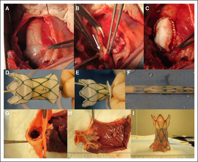
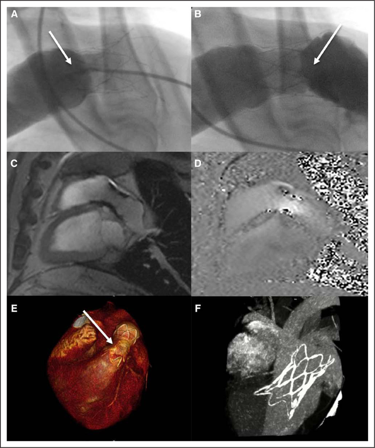
Methods and results: Thirteen sheep underwent baseline cardiac magnetic resonance imaging, surgical pulmonary valvectomy, and transannular patch repair. One month after transannular patch repair, the hTPV was implanted, followed by serial magnetic resonance imaging and computed tomography imaging at 1, 5, and 8 month(s). hTPV implantation was successful in 11 animals (85%). There were 2 procedural deaths related to ventricular fibrillation. Seven animals survived the entire follow-up protocol, 5 with functioning hTPV devices. Two animals had occlusion of hTPV with aneurysm of main pulmonary artery. A strong decline in pulmonary regurgitant fraction was observed after hTPV implantation (40.5% versus 8.3%; P=0.011). Right ventricular end diastolic volume increased by 49.4% after transannular patch repair (62.3-93.1 mL/m2; P=0.028) but was reversed to baseline values after hTPV implantation (to 65.1 mL/m2 at 8 months, P=0.045). Both right ventricular ejection fraction and left ventricular ejection fraction were preserved after hTPV implantation.

Conclusions: hTPV implantation is feasible, significantly reduces pulmonary regurgitant fraction, facilitates right ventricular volume improvements, and preserves biventricular function in an ovine model of chronic pulmonary insufficiency. This percutaneous strategy could potentially offer an alternative for standard surgical pulmonary valve replacement in dilated right ventricular outflow tracts, permitting lower risk, nonsurgical pulmonary valve replacement in previously prohibitive anatomies.

Keywords: cardiac catheterization; computed tomography; heart valve prosthesis implantation; magnetic resonance imaging; pulmonary valve regurgitation; tetralogy of Fallot.


Conflict of interest statement

Disclosures

Dr Gillespie serves as a consultant to Medtronic, the manufacturer of the hTPV device. Dr Litt receives research funding from Siemens Healthcare for unrelated projects. The other authors report no conflicts.

Figures

Figure 1
Figure 1
Photographs of the surgical creation of the ovine pulmonary insufficiency model, followed by transannular patching and Harmony transcatheter pulmonary valve (hTPV) implantation. Intraoperative photographs show the RVOT prevalvectomy (A) and the opened pulmonary artery (PA) with one leaflet of the pulmonary valve being excised (B, arrow). C, Intraoperative picture of transannular patch repair. D, The hTPV in expanded state. E, The hTPV is being loaded into the delivery catheter. F, The hTPV is entirely collapsed into the delivery catheter. G, Picture of the distal PA in the excised heart, showing the hTPV firmly fixed in the PA. H, Photograph shows the device appropriately positioned into the RVOT in situ. I, The hTPV was eventually excised, and valve leaflets were examined. There was no device fracture or erosion. RVOT indicates right ventricular outflow tract.
Figure 2
Figure 2
Typical RVOT anatomy at 1-month post-transannular patch repair and pre- Harmony transcatheter pulmonary valve implantation, presented with cine magnetic resonance imaging (A), phase-contrast magnetic resonance imaging (B), and computed tomography (C). The angiogram shows the dimensions of the right ventricular outflow tract (RVOT) (D). For all animals, the diameter of the pulmonary annulus after transannular patch repair ranged between 29 and 32 mm in systole. LV indicates left ventricle; and RV, right ventricle.
Figure 3
Figure 3
Imaging of the right ventricular outflow tract after successful Harmony transcatheter pulmonary valve (hTPV) implantation. The angiograms show excellent proximal (A) and distal (B) seal (arrows). Sagittal magnetic resonance imaging (C) and velocity mapping (D) reveal no significant (para)valvular leaking. The hTPV is well positioned (arrow), stable, and no stent fractures were observed (E and F, computed tomography imaging).
Figure 4
Figure 4
Figure shows means and SE bars. 1: 1 month after Harmony transcatheter pulmonary valve (hTPV) implantation, 5: 3 to 5 months after hTPV implantation, 8: 6 to 8 months after hTPV implantation. PRF indicates pulmonary regurgitant fraction.
Figure 5
Figure 5
Indexed right ventricular volumes at each time point. Figure shows means and SE bars. 1: 1 month after Harmony transcatheter pulmonary valve (hTPV) implantation, 5: 3 to 5 months after hTPV implantation, 8: 6 to 8 months after hTPV implantation. EDV indicates end diastolic volume; and ESV, end systolic volume.
Figure 6
Figure 6
Ejection fraction for the right ventricle (RV) and left ventricle (LV). Figure shows means and SE bars. 1: 1 month after Harmony transcatheter pulmonary valve (hTPV) implantation, 5: 3 to 5 months after hTPV implantation, 8: 6 to 8 months after hTPV implantation. LVEF indicates left ventricular ejection fraction; and RVEF, right ventricular ejection fraction.
Figure 7
Figure 7
Four-dimensional flow visualization showing flow dynamics in the right ventricular (RV) outflow tract at baseline (A and B), postvalvectomy (C and D) and 8 months after Harmony transcatheter pulmonary valve (hTPV) implantation (E and F). Cardiac phases are indicated late diastole (A, C, E) and late systole (B, D, F). Severe regurgitation crashing into the RV wall was observed during diastole after valvectomy (C, thick arrow), compared with almost zero regurgitation at baseline (A, dotted arrow) and little regurgitation at 8-month post-hTPV (E, dashed arrow). Velocity scale score in m/s. LPA indicates left pulmonary artery; MPA, main pulmonary artery; RPA, right pulmonary artery; and RV, right ventricular.
Figure 8
Figure 8
Harmony transcatheter pulmonary valve (hTPV) with occluded lumen and aneurysm formation in 1 animal. A, Angiogram showing perivalvular channels (arrow); B, cardiac magnetic resonance imaging displaying the right ventricular outflow tract with visible flow outside the stent, as indicated by the thick arrow; C, computed tomography showing perivalvular channels around the hTPV (arrow); D, postmortem examination of the stent showing occlusion of the valve, the arrow points out the occluded leaflets of the hTPV.

References

    1. Apitz C, Webb GD, Redington AN. Tetralogy of Fallot. Lancet 2009;374:1462–1471. doi: 10.1016/S0140-6736(09)60657-7. - DOI - PubMed
    1. Reller MD, Strickland MJ, Riehle-Colarusso T, Mahle WT, Correa A. Prevalence of congenital heart defects in metropolitan Atlanta, 1998–2005. J Pediatr 2008;153:807–813. doi: 10.1016/j.jpeds.2008.05.059. - DOI - PMC - PubMed
    1. Luijten LW, van den Bosch E, Duppen N, Tanke R, Roos-Hesselink J, Nijveld A, van Dijk A, Bogers AJ, van Domburg R, Helbing WA. Long-term outcomes of transatrial-transpulmonary repair of tetralogy of Fallot. Eur J Cardiothorac Surg 2015;47:527–534. doi: 10.1093/ejcts/ezu182. - DOI - PubMed
    1. Al Habib HF, Jacobs JP, Mavroudis C, Tchervenkov CI, O’Brien SM, Mohammadi S, Jacobs ML. Contemporary patterns of management of tetralogy of Fallot: data from the Society of Thoracic Surgeons Database. Ann Thorac Surg 2010;90:813–819, discussion 819. doi: 10.1016/j.athoracsur.2010.03.110. - DOI - PubMed
    1. Ferraz Cavalcanti PE, Sá MP, Santos CA, Esmeraldo IM, de Escobar RR, de Menezes AM, de Azevedo OM Jr, de Vasconcelos Silva FP, Lins RF, Lima Rde C. Pulmonary valve replacement after operative repair of tetralogy of Fallot: meta-analysis and meta-regression of 3,118 patients from 48 studies. J Am Coll Cardiol 2013;62:2227–2243. doi: 10.1016/j.jacc.2013.04.107. - DOI - PubMed