. 2016 Nov 4;291(45):23516-23531.
doi: 10.1074/jbc.M116.735399. Epub 2016 Sep 23.

A Novel Conserved Domain Mediates Dimerization of Protein Kinase D (PKD) Isoforms: DIMERIZATION IS ESSENTIAL FOR PKD-DEPENDENT REGULATION OF SECRETION AND INNATE IMMUNITY

Affiliations

A Novel Conserved Domain Mediates Dimerization of Protein Kinase D (PKD) Isoforms: DIMERIZATION IS ESSENTIAL FOR PKD-DEPENDENT REGULATION OF SECRETION AND INNATE IMMUNITY

Clara Aicart-Ramos et al. J Biol Chem. .

Abstract

Protein kinase D (PKD) isoforms are protein kinase C effectors in signaling pathways regulated by diacylglycerol. Important physiological processes (including secretion, immune responses, motility, and transcription) are placed under diacylglycerol control by the distinctive substrate specificity and subcellular distribution of PKDs. Potentially, broadly co-expressed PKD polypeptides may interact to generate homo- or heteromultimeric regulatory complexes. However, the frequency, molecular basis, regulatory significance, and physiological relevance of stable PKD-PKD interactions are largely unknown. Here, we demonstrate that mammalian PKDs 1-3 and the prototypical Caenorhabditis elegans PKD, DKF-2A, are exclusively (homo- or hetero-) dimers in cell extracts and intact cells. We discovered and characterized a novel, highly conserved N-terminal domain, comprising 92 amino acids, which mediates dimerization of PKD1, PKD2, and PKD3 monomers. A similar domain directs DKF-2A homodimerization. Dimerization occurred independently of properties of the regulatory and kinase domains of PKDs. Disruption of PKD dimerization abrogates secretion of PAUF, a protein carried in small trans-Golgi network-derived vesicles. In addition, disruption of DKF-2A homodimerization in C. elegans intestine impaired and degraded the immune defense of the intact animal against an ingested bacterial pathogen. Finally, dimerization was indispensable for the strong, dominant negative effect of catalytically inactive PKDs. Overall, the structural integrity and function of the novel dimerization domain are essential for PKD-mediated regulation of a key aspect of cell physiology, secretion, and innate immunity in vivo.

Keywords: Caenorhabditis elegans (C. elegans); heterodimer; homodimer; innate immunity; native Mr; oligomerization domain; protein kinase D (PKD); secretion; signal transduction.


Figures

FIGURE 1.
FIGURE 1.
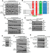
A novel N-terminal structural module in C. elegans DKF-2A mediates PKD-PKD interactions that generate a homomultimeric kinase. A, HEK293 cells were co-transfected with transgenes encoding HA or FLAG-tagged DKF-2A (designated 2A), DKF-2B (2B), or constitutively active DKF-2A EE (2A*). After 48 h, the cells were lysed, and PKDs were precipitated from cell extracts by adding anti-HA IgGs and protein G-Sepharose 4B beads. Precipitated and co-precipitated proteins were analyzed by SDS-PAGE and Western blotting, using anti-HA and anti-FLAG IgGs, respectively. The same IgGs detected epitope-tagged PKDs in blots of cell extracts. Tubulin was monitored as a loading control. B, a schematic diagram depicts mutant DKF-2A proteins and DKF-2A domains that were assayed for oligomerization activity. In C–G, transfection, lysis, immunoprecipitation, and Western blot analysis were performed as indicated in A and “Experimental Procedures.” C, FLAG-DKF-2A or a FLAG-DKF-2A truncation mutant was co-expressed with HA-DKF-2A. D, FLAG-DKF-2A or a FLAG-tagged DKF-2A internal deletion mutant was co-expressed with HA-DKF-2A. E, a DKF-2A 311–545-mCherry fusion protein, which contains the C1a and C1b DAG binding sites, and HA-DKF-2A were co-expressed. The cells were treated with 1 μm PMA or vehicle for 20 min prior to lysis. F, FLAG-DKF-2A or a FLAG-DKF-2A truncation mutant was co-expressed with the DKF-2A 1–319-GFP fusion protein. G, cells were co-transfected with a fixed amount of HA-DKF-2A transgene and increasing amounts of DKF-2A 1–319-mCherry fusion transgene. IB, immunoblot; IP, immunoprecipitation; PH, pleckstrin homology.
FIGURE 2.
FIGURE 2.
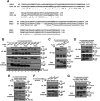
Human PKDs assemble into homo- and hetero-oligomers; a conserved domain mediates PKD multimerization. A, human PKD2 and PKD1 have a conserved domain that shares substantial amino acid sequence identity (*) and similarity (:) with the DKF-2A OD. B–G, transfection, lysis, immunoprecipitation and Western blot analysis were performed as indicated in Fig. 1A. B, pairs of HA and FLAG-tagged PKD isoforms or empty vector (−) were co-expressed in HEK293 cells. C, FLAG-PKD2 or a FLAG-PKD2 N-terminal truncation mutant was co-expressed with HA-PKD2. D, HA-PKD1, HA-PKD2, or HA-PKD3 was co-expressed with a chimeric protein containing amino acids 1–146 of PKD2 fused with mCherry. E, HA-PKD1, HA-PKD2, or HA-PKD3 was co-expressed with a FLAG-tagged protein containing amino acids 1–142 of PKD3. F, FLAG-PKD1, FLAG-PKD2, or FLAG-PKD1 EE was co-expressed with an internal deletion mutant of HA-PKD1 EE (PKD1 EE Δ50–125). G, a protein that contains the C1a and C1b binding domains of PKD2 (amino acids 128–328) fused to mCherry was co-expressed with HA-tagged PKD1, PKD2, or PKD3. IB, immunoblot; IP, immunoprecipitation.
FIGURE 3.
FIGURE 3.
PKDs oligomerize in intact cells. A, cells co-expressing HA-DKF-2A and FLAG-DKF-2A were incubated with the indicated concentrations of DFDNB or DSS cross-linker or vehicle. Subsequently cross-linking was quenched, the cells were lysed, and PKD oligomerization was monitored by SDS-PAGE and Western blot analysis as described under “Experimental Procedures.” Positions of monomeric DKF-2A (M) and high molecular weight DKF-2A oligomers (O) are marked. B, the Western blot in A was stripped and reprobed with IgGs directed against endogenous PKD2, PKCα, and PKCδ. C, cells expressing HA-DKF-2A and FLAG-DKF-2A were treated with reversible cross-linker DSP as described in A. Replicate samples of Triton X-100-soluble proteins were denatured in SDS-PAGE loading buffer containing or lacking 5% 2-mercaptoethanol. Oligomerization was monitored as described in A. An asterisk marks the position of a nonspecific band. D, the Western blot in C was stripped and reprobed with IgGs directed against endogenous PKD2. PKD2 monomers (M) and oligomers (O) are indicated. E, cells expressing HA-PKD1 Δ50–125 were incubated in the absence or presence of 0.3 mm DFDNB. Extracted proteins were analyzed as described for A. The position of monomeric PKD1 Δ50–125 (M) is marked; no oligomers were detected. IB, immunoblot.
FIGURE 4.
FIGURE 4.
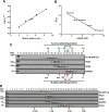
DKF-2A and PKDs 1 and 2 are dimeric proteins. A, a 1 × 30-cm Superose 6 gel filtration column was calibrated with purified proteins with established Stokes radii: thyroglobulin (8.5 nm), apoferritin (6.2 nm), ferritin (6.2 nm), catalase (5.2 nm), aldolase (4.8 nm), ADH (4.5 nm), albumin (3.6 nm), and myoglobin (2.0 nm). Kav = VeVo/VtVo, where Ve is the elution volume, Vo is the void volume, and Vt is the total column volume. The log-linear relationship of the plot enables accurate interpolation of experimental Rs values by using Graph Pad Prism 6 linear regression software. The details are provided under “Experimental Procedures.” B, a 5–30% sucrose density gradient was calibrated for determination of S20,W values of PKDs. Purified proteins with established S20,W values, catalase (11.3), aldolase (7.4), ADH (7.3), albumin (4.6), and myoglobin (1.9), were centrifuged at 36,000 rpm for 26 h at 5 °C in an SW41 Ti rotor. Proteins in fractions collected from the gradient were characterized by SDS-PAGE and Coomassie Blue staining. Graph Pad Prism 6 linear regression software was used to determine experimental S20,W values (see “Experimental Procedures: and Refs. and 35). C, detergent-soluble proteins were extracted from cells expressing FLAG-DKF-2A and HA-DKF-2A. Proteins were fractionated according to Rs by gel filtration on Superose 6. DKF-2A and endogenous PKD1, PKD2, and GAPDH were detected by assaying aliquots of column fractions via Western immunoblotting. Yellow dots show positions of protein peaks. Experimentally determined Rs values are listed in Table 1. Native molecular weights of calibrating proteins (red arrows) are shown to illustrate that PKDs appear to be large oligomers in the absence of information about S20,W values. Only relevant fractions are shown; Vo was collected in fractions 1–8. T indicates the signal obtained from a sample of total protein extract. D, a sample of lysate described in C was fractionated in a sucrose gradient. Fractions were collected and analyzed as described under “Experimental Procedures.” Protein peaks are marked with yellow dots. Peaks of calibrating proteins are indicated with red arrows. Experimentally determined S20,W values are given in Table 1.
FIGURE 5.
FIGURE 5.
Dimerization is an intrinsic property of PKDs. A, GST-His6-DKF-2A was expressed in Sf9 cells infected with recombinant baculovirus and then purified by affinity chromatography on a column of Ni2+-NTA-agarose (see “Experimental Procedures”). Samples of total detergent-soluble protein (ST), flow through (FT) proteins, and proteins collected during washing of the resin and elution of GST-His6-DKF-2A with 0.25 m imidazole were analyzed by SDS-PAGE and staining with Coomassie Blue. B, after subsequent affinity chromatography on GSH-Sepharose 4B and cleavage of the GST-His6 tag by thrombin, DKF-2A was further purified and characterized by FPLC gel filtration on Superose 6. Aliquots of fractions were analyzed by SDS-PAGE, silver staining (B), and Western blotting with anti-DKF-2A IgGs (C). D, His-PKD2 was expressed, purified on Ni2+-NTA-agarose, and characterized as described above. Western immunoblot analyses of aliquots of fractions collected from the Superose 6 column are shown.
FIGURE 6.
FIGURE 6.
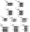
Disruption of dimerization blocks PKD-mediated secretion. A, cells co-expressing PAUF-Myc and either HA-PKD1, HA-PKD1 KD, or HA-PKD EE were assayed for accumulation of immature PAUF in cell extracts and secreted PAUF in serum-free OptiMEM medium as described under “Experimental Procedures.” A PKD-selective inhibitor NX-6 was added as indicated. Immature and secreted PAUF were detected by Western immunoblot analysis using anti-Myc IgGs. B, triply transfected HEK293 cells co-expressing PAUF-Myc and HA-PKD1 EE, along with either the PKD2 OD (PKD2 1–146-mCherry), the PKD3 OD (FLAG-PKD3 1–142), or empty vector were assayed for PAUF production and secretion. C, cells co-expressing PAUF-Myc and HA-PKD1, along with either the PKD2 OD, the PKD3 OD, or empty vector were assayed for PAUF secretion. D, cells co-expressing PAUF-Myc and HA-PKD2 EE, along with either the PKD2 OD or empty vector were assayed for PAUF secretion. E, cells co-expressing PAUF-Myc and either HA-PKD1 KD, HA-PKD2 KD, or empty vector were assayed for PAUF secretion. PMA was added to OptiMEM secretion medium. F, cells co-expressing PAUF-Myc and either HA-PKD1 KD, HA-PKD1 KD Δ50–125, PKD2 1–146-mCherry, or empty vector were assayed for PAUF secretion. PMA was added to OptiMEM medium. G, cloned HEK293 cells stably expressing a PAUF-Myc transgene were transiently transfected with empty vector or transgenes encoding HA-PKD1 KD or PKD2 1–146-mCherry. The cells were assayed for PAUF secretion as above. H, extracts of cells co-expressing GFP-PI4KIIIβ and either FLAG-PKD2 or a dimerization-defective FLAG-PKD2 mutant (Δ66 or Δ138) were assayed for PI4KIIIβ phosphorylation. GFP-PI4KIIIβ was isolated from cell extracts by immunoprecipitation with anti-GFP IgG; Western immunoblot analysis was performed using anti-phospho-PKD substrate IgGs as primary antibody. The cells were treated with 1 μm PMA or vehicle for 20 min prior to lysis. I, cells expressing both GFP-PI4KIIIβ and HA-DKF-2A were co-transfected with a transgene encoding DKF-2A 1–319-mCherry (dimerization domain) or empty vector and were assayed for PI4KIIIβ phosphorylation as described in H above. The cells were treated with 1 μm PMA or vehicle for 20 min prior to lysis. J, cells expressing GFP-PI4KIIIβ were co-transfected with transgenes encoding HA-DKF-2A EE and DKF-2A 1–319-mCherry (dimerization domain) or empty vector, as indicated. Phosphorylation of PI4KIIIβ was assayed via Western immunoblotting as described in H above. All experiments were repeated three times; each replication yielded similar results. IB, immunoblot; IP, immunoprecipitation.
FIGURE 7.
FIGURE 7.
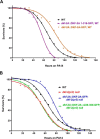
Disruption of DKF-2A dimerization impairs innate immunity. A, survival curves for WT and transgenic (dkf-2A::DKF-2A 1–319-GFP, WT background and dkf-2A::DKF-2A-GFP, WT background) C. elegans fed with pathogenic PA14. B, survival curves for WT, dkf-2(pr3) null and transgenic (dkf-2A::DKF-2A Δ228–300-GFP, dkf-2(pr3) and dkf-2A::DKF-2A-GFP, dkf-2(pr3) C. elegans fed with PA14. All assays were replicated three times with similar results; typical data are shown.

Similar articles

Cited by

References

    1. Newton A. C. (2010) Protein kinase C: poised to signal. Am. J. Physiol. Endocrinol. Metab. 298, E395–E402 - PMC - PubMed
    1. Steinberg S. F. (2008) Structural basis of protein kinase C isoform function. Physiol. Rev. 88, 1341–1378 - PMC - PubMed
    1. Fu Y., and Rubin C. S. (2011) Protein kinase D: coupling extracellular stimuli to the regulation of cell physiology. EMBO Rep. 12, 785–796 - PMC - PubMed
    1. Rozengurt E. (2011) Protein kinase D signaling: multiple biological functions in health and disease. Physiology 26, 23–33 - PMC - PubMed
    1. Ellwanger K., and Hausser A. (2013) Physiological functions of protein kinase D in vivo. IUBMB Life 65, 98–107 - PubMed

LinkOut - more resources