. 2016 Nov 4;291(45):23693-23703.
doi: 10.1074/jbc.M116.749341. Epub 2016 Sep 23.

Polo-like Kinase 1 Regulates Vimentin Phosphorylation at Ser-56 and Contraction in Smooth Muscle

Affiliations

Polo-like Kinase 1 Regulates Vimentin Phosphorylation at Ser-56 and Contraction in Smooth Muscle

Jia Li et al. J Biol Chem. .

Abstract

Polo-like kinase 1 (Plk1) is a serine/threonine-protein kinase that has been implicated in mitosis, cytokinesis, and smooth muscle cell proliferation. The role of Plk1 in smooth muscle contraction has not been investigated. Here, stimulation with acetylcholine induced Plk1 phosphorylation at Thr-210 (an indication of Plk1 activation) in smooth muscle. Contractile stimulation also activated Plk1 in live smooth muscle cells as evidenced by changes in fluorescence resonance energy transfer signal of a Plk1 sensor. Moreover, knockdown of Plk1 in smooth muscle attenuated force development. Smooth muscle conditional knock-out of Plk1 also diminished contraction of mouse tracheal rings. Plk1 knockdown inhibited acetylcholine-induced vimentin phosphorylation at Ser-56 without affecting myosin light chain phosphorylation. Expression of T210A Plk1 inhibited the agonist-induced vimentin phosphorylation at Ser-56 and contraction in smooth muscle. However, myosin light chain phosphorylation was not affected by T210A Plk1. Ste20-like kinase (SLK) is a serine/threonine-protein kinase that has been implicated in spindle orientation and microtubule organization during mitosis. In this study knockdown of SLK inhibited Plk1 phosphorylation at Thr-210 and activation. Finally, asthma is characterized by airway hyperresponsiveness, which largely stems from airway smooth muscle hyperreactivity. Here, smooth muscle conditional knock-out of Plk1 attenuated airway resistance and airway smooth muscle hyperreactivity in a murine model of asthma. Taken together, these findings suggest that Plk1 regulates smooth muscle contraction by modulating vimentin phosphorylation at Ser-56. Plk1 activation is regulated by SLK during contractile activation. Plk1 contributes to the pathogenesis of asthma.

Keywords: cytoskeleton; excitation-contraction coupling (E-C coupling); intermediate filament; phosphorylation; signal transduction; smooth muscle.


Figures

FIGURE 1.
FIGURE 1.
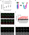
Phosphorylation and activation of Plk1 in smooth muscle cells. A, smooth muscle cells were stimulated with ACh (10−4 m) for 1–10 min or left unstimulated. Plk1 phosphorylation at Thr-210 was evaluated by immunoblot analysis. Data are the mean values of experiments from five batches of cell culture. Error bars indicate S.D. B, schematic diagram of Plk1 biosensor. In inactive status, PBD binds to the catalytic domain forming a closed conformation. The closed conformation renders N-terminal DsRed and C-terminal GFP proximity, leading to high levels of FRET and lower GFP/DsRed ratios. Upon activation, Plk1 becomes an open conformation. The distance between the N-terminal DsRed and C-terminal GFP increases, which leads to low FRET signal and higher GFP/DsRed ratios. The open conformation facilitates the recruitment of substrate to the catalytic domain. C, representative images illustrating the effects of ACh stimulation (10−4 m) on emission of GFP and DsRed in HASM cells. The insets are the 4× magnification of the selected areas. WT, wild type Plk1; T210A, T210A Plk1; ”, seconds. D, stimulation with ACh increases the activity of WT Plk1 but not T210A Plk1. Ratios of GFP/DsRed fluorescence were determined for HASM cells in response to ACh stimulation (10−4 m). Increases in GFP/DsRed ratios indicate higher Plk1 activity. Data are the means ± S.D. (n = 15 cells from 4 batches of cell culture for WT Plk1 and 12 cells from 4 batches of cell culture for T210A Plk1). E, the GFP and DsRed emissions of cells expressing GFP-tagged Plk1 were evaluated using a confocal microscope. GFP emission was very high, whereas DsRed signal was not detected. In addition, ACh stimulation did not affect GFP/DsRed emission. Because no DsRed (RED) emission was detected, we failed to calculate the ratio of GFP/DsRed (n = 12 cells from 4 batches of cell culture). DIC, differential interference contrast. F, the emissions of GFP and DsRed of cells expressing DsRed-tagged construct were assessed. Neither GFP nor DsRed was found under the experimental conditions. ACh treatment did not affect the emission of GFP and DsRed. No GFP/DsRed ratios were calculated because of lack of fluorescent signals (n = 14 cells from 4 batches of cell culture). Scale bar: 10 μm.
FIGURE 2.
FIGURE 2.
Plk1 is necessary for smooth muscle contraction. A, human bronchial rings were transduced with lentiviruses encoding control shRNA or Plk1 shRNA. These tissues were then incubated in the serum-free medium for 3 days. Immunoblot analysis was used to assess protein expression in tissues. UI, uninfected. *, significantly lower protein ratios of Plk1/GAPDH in tissues transduced with virus encoding Plk1 shRNA than in uninfected tissues and tissues expressing control shRNA (p < 0.05). Data are the mean values of experiments from three donors. Error bars indicate S.D. B, contraction of human bronchial rings to ACh was evaluated, after which they were transduced with lentiviruses as described above. Contractile responses were compared before and after incubation. *, significantly lower contractile force in bronchial rings treated with Plk1 shRNA as compared with uninfected tissues or tissues infected with viruses encoding control shRNA (p < 0.05). Data are the mean values of 6 samples from 3 donors. Error bars indicate S.D. C, tracheal rings of Plk1smko mice and Plk1−lox mice were treated with difference concentration of ACh. Dose response of these rings was then evaluated. Contractile force is normalized to maximal force induced by 10−3 m ACh. Data are the mean values of experiments from six mice/each group. Error bars indicate S.D. *, significantly lower active force in Plk1smko mice than in Plk1−lox at corresponding doses (p < 0.05). Inserted immunoblots show Plk1 protein expression in airway smooth muscle cells from Plk1−lox and Plk1smko mice. Blots are representative of four identical experiments. D, tracheal rings of wild type mice were precontracted with 10 μm ACh. Different concentrations of BI6727 were then imposed to assess the relaxation response. Treatment with the pharmacological inhibitor induced relaxation of tracheal segments precontracted by ACh. Data are the mean values of experiments from four mice. Error bars indicate S.D. *, p < 0.05 versus contraction before the addition of BI6727.
FIGURE 3.
FIGURE 3.
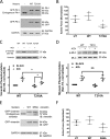
Knockdown of Plk1 attenuates the ACh-induced vimentin phosphorylation at Ser-56 without affecting myosin light chain phosphorylation at Ser-19. A, smooth muscle cells expressing control shRNA or Plk1 shRNA were stimulated with ACh (10−4 m, 5 min) or left unstimulated. Vimentin phosphorylation at Ser-56 was evaluated by immunoblot analysis. Data are the mean values of four batches of cell culture. Error bars indicate S.D. (*, p < 0.05). B, purified active Plk1 (20 ng) and 1 μg of wild type vimentin or mutant S56A vimentin were placed in kinase buffer. Vimentin phosphorylation at Ser-56 was determined by immunoblot analysis 30 min after the initiation of the reaction. Vimentin phosphorylation catalyzed by Plk1 was normalized to the level of vimentin phosphorylation in the absence of Plk1. Data are the mean values of four in vitro biochemical experiments. Error bars indicate S.D. (*, p < 0.05). C, myosin light chain (MLC) phosphorylation at Ser-19 in cells transduced with lentivirus encoding control or Plk1 shRNA was assessed by immunoblot analysis. Myosin phosphorylation was similar in cells expressing control shRNA or Plk1 shRNA (NS, not significant). Data are the mean values of four batches of cell culture. Error bars indicate S.D.
FIGURE 4.
FIGURE 4.
Roles of Plk1 phosphorylation at Thr-210 and vimentin Ser-56 phosphorylation in smooth muscle contraction. A, representative immunoblots (IB) illustrating the expression of WT Plk1 or T210A Plk1 mutant. Extracts of human bronchial tissues transduced with plasmids encoding WT Plk1 or T210A Plk1 mutant were immunoblotted with antibodies against Plk1, GFP, or GAPDH. GFP-Plk1 was detected in the extracts of tissues transduced with plasmid for WT or T210A Plk1. Blots are representative of experiments from three donors. B, contraction of human bronchial rings was evaluated, after which they were transduced with the plasmids as described under “Experimental Procedures.” Contractile responses were compared before and after incubation. *, significantly lower contractile force in bronchial rings expressing T210A Plk1 as compared with tissues transfected with WT Plk1 or untransfected (UT) tissues (p < 0.05). Data are the mean values of experiments from three donors. Error bars indicate S.D. C, cells expressing WT or T210A Plk1 were stimulated with ACh (10−4 m, 5 min) or left unstimulated. Vimentin phosphorylation at Ser-56 in the cells was evaluated using immunoblot analysis. Data are the mean values of experiments from four batches of cell culture. Error bars indicate S.D. (*, p > 0.05). D, myosin light chain (MLC) phosphorylation at Ser-19 in cells expressing WT or T210A Plk1 was assessed by immunoblot analysis. Myosin phosphorylation was similar in cells expressing WT or T210A Plk1 (NS, not significant). Data are the mean values of experiments from four batches of cell culture. Error bars represent S.D. E, representative immunoblots showing the expression of WT and S56A vimentin. Extracts of human bronchial tissues transduced with plasmids encoding WT or S56A vimentin were probed with antibodies against vimentin, GFP, or GAPDH. Blots are representative of experiments from three donors. F, contraction of tissues expressing S56A vimentin is reduced as compared with tissues treated with WT vimentin or UT tissues. Data are the mean values of experiments from three donors. Error bars indicate S.D.
FIGURE 5.
FIGURE 5.
Knockdown of SLK inhibits the ACh-induced phosphorylation and activation of Plk1. A, extracts of smooth muscle cells transfected with control siRNA or SLK siRNA for 2 days were immunoblotted with antibodies against SLK or GAPDH. *, significantly lower protein ratios of SLK/GAPDH in cells treated with SLK siRNA than in cells treated with control siRNA (p < 0.05). Data are the mean values of experiments from five batches of cell culture. Error bars indicate S.D. B, smooth muscle cells transfected with control or SLK siRNA for days were stimulated with ACh (10−4 m, 5 min) or left unstimulated. Plk1 phosphorylation at Thr-210 was evaluated by immunoblot analysis. Data are the mean values of experiments from four batches of cell culture. Error bars indicate S.D. (*, p < 0.05). C, cells treated with control or Plk1 siRNA were transfected with plasmids for Plk1 sensor. These cells were stimulated with ACh (10−4 m) 2 days after transfection. The fluorescence of GFP/DsRed was captured live using a laser confocal microscope. The ratios of GFP/DsRed fluorescence were determined using the method described under “Experimental Procedures.” Values represent the means ± S.D. (n = 14 cells from 4 batches of each cell culture). *, Significantly different at each time point between cells treated with control siRNA and SLK KD cells (p < 0.05).
FIGURE 6.
FIGURE 6.
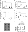
Knock-out or inhibition of Plk1 diminishes airway resistance and contractile response of tracheal rings from HDM-exposed mice. A, Plk1−lox and Plk1smko mice were exposed to HDM for 6 weeks. Airway resistance (RAW) in response to inhalation of 80 mg/kg MCh was then measured. The data represent the means ± S.D. (*, p < 0.05). **, significantly lower airway resistance in Plk1smko mice treated with PBS than in Plk1−lox mice treated with PBS (p < 0.05). n = 6–8 mice/each group. B, the contractile response of tracheal rings from these mice was determined by using an in vitro organ bath system. Contractile force is normalized to the mean maximal force of rings from HDM-treated Plk1−lox mice. The data represent the means ± S.D. (*, p < 0.05). **, significantly lower tracheal contraction of Plk1smko mice treated with PBS than Plk1−lox mice treated with PBS (p < 0.05). n = 6–8 mice/each group. C, C57BL/6J mice were exposed to HDM in the presence or absence of the Plk1 inhibitor BI6727 as described under “Experimental Procedures.” Intranasal instillation of BI6727 inhibits RAW in mice treated with HDM. The data represent the means ± S.D. (*, p < 0.05, n = 4–6 mice/group). D, treatment with the Plk1 inhibitor BI6727 attenuates HDM-sensitized tracheal contraction ex vivo. Contractile force is normalized to maximal force of rings from HDM- and vehicle-treated mice. Error bars indicate S.D. (*, p < 0.05, n = 4–5 mice/group). E, the size of lungs from Plk1smko mice is similar to that from Plk1−lox mice. Shown are images of lungs from 3 Plk1−lox mice and 3 Plk1smko mice. Scale bar, 1 cm. F, blots of tracheal smooth muscle tissues from Plk1−lox mice and Plk1smko mice were probed with antibodies against smooth muscle myosin heavy chain 11 (MYH11), α-actin. and GAPDH. Protein ratios of MYH11/GAPDH and α-actin/GAPDH from Plk1smko mice are normalized to those from Plk1−lox mice. The data represent the means ± S.D. (NS, not significant, n = 3 mice/group).
FIGURE 7.
FIGURE 7.
Role and regulation of Plk1 in smooth muscle. Contractile stimulation induces phosphorylation and activation of Plk1 mediating vimentin phosphorylation at Ser-56, which may facilitate intracellular/intercellular force transduction and smooth muscle contraction. Plk1 activation is mediated by SLK.

Similar articles

Cited by

References

    1. Garrod D. R., Merritt A. J., and Nie Z. (2002) Desmosomal adhesion: structural basis, molecular mechanism, and regulation. Mol. Membr. Biol. 19, 81–94 - PubMed
    1. Small J. V., and Gimona M. (1998) The cytoskeleton of the vertebrate smooth muscle cell. Acta Physiol. Scand. 164, 341–348 - PubMed
    1. Wang R., Li Q., and Tang D. D. (2006) Role of vimentin in smooth muscle force development. Am. J. Physiol. Cell Physiol. 291, C483–C489 - PMC - PubMed
    1. Wang R., Li Q. F., Anfinogenova Y., and Tang D. D. (2007) Dissociation of Crk-associated substrate from the vimentin network is regulated by p21-activated kinase on ACh activation of airway smooth muscle. Am. J. Physiol. Lung Cell Mol. Physiol. 292, L240–L248 - PMC - PubMed
    1. Tang D. D. (2008) Invited review: intermediate filaments in smooth muscle. Am. J. Physiol. Cell Physiol 294, C869–C878 - PMC - PubMed