Review
. 2016 Aug;43(4):464-75.
doi: 10.1053/j.seminoncol.2016.06.004. Epub 2016 Jun 15.

Promise and limits of the CellSearch platform for evaluating pharmacodynamics in circulating tumor cells

Affiliations
Review

Promise and limits of the CellSearch platform for evaluating pharmacodynamics in circulating tumor cells

Lihua Wang et al. Semin Oncol. 2016 Aug.

Abstract

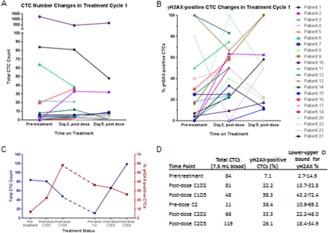
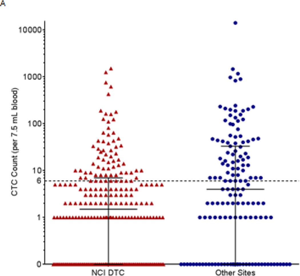
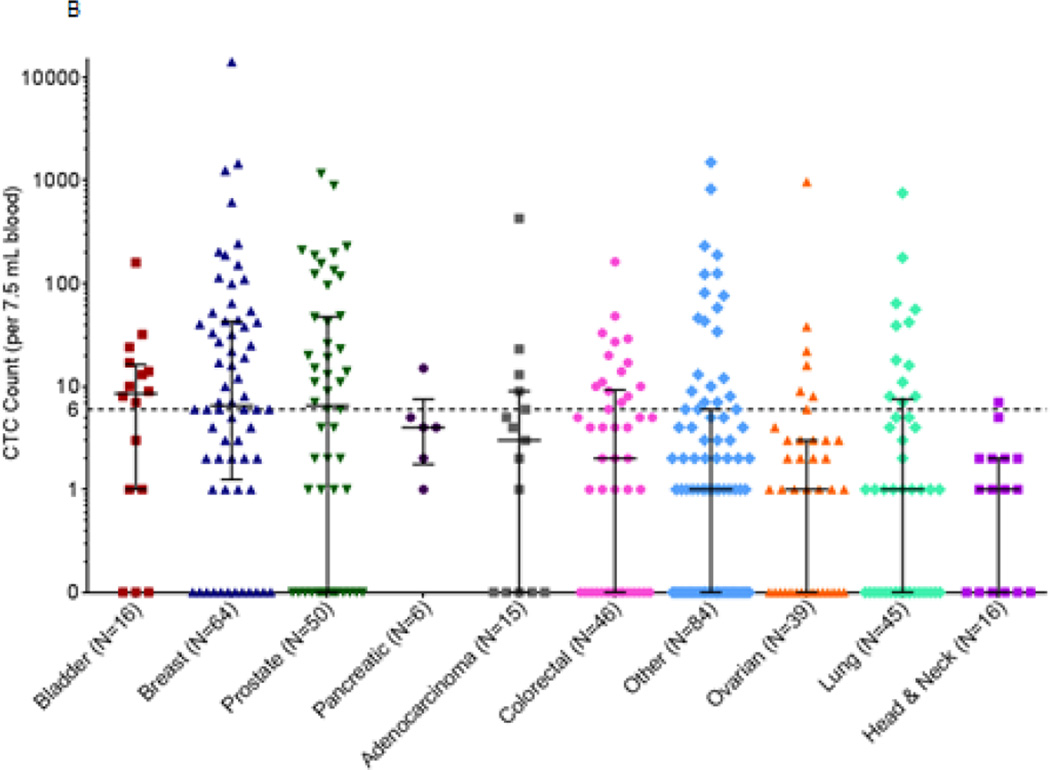
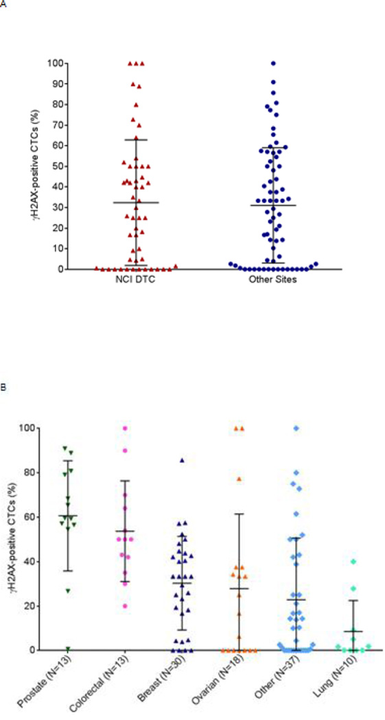
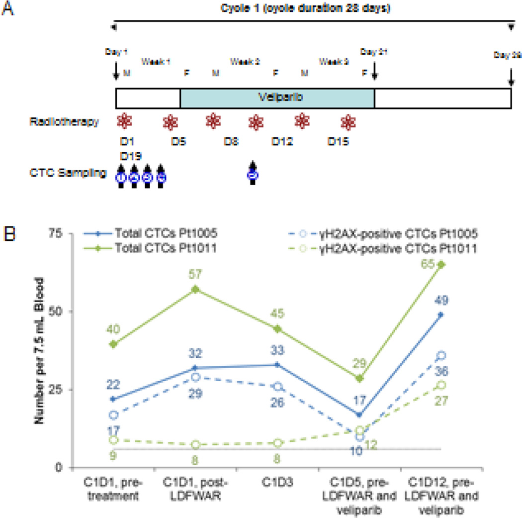
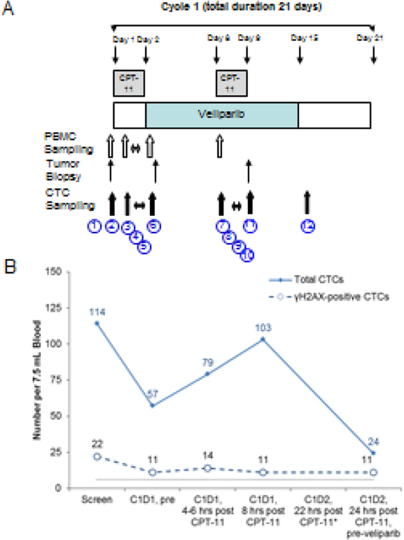
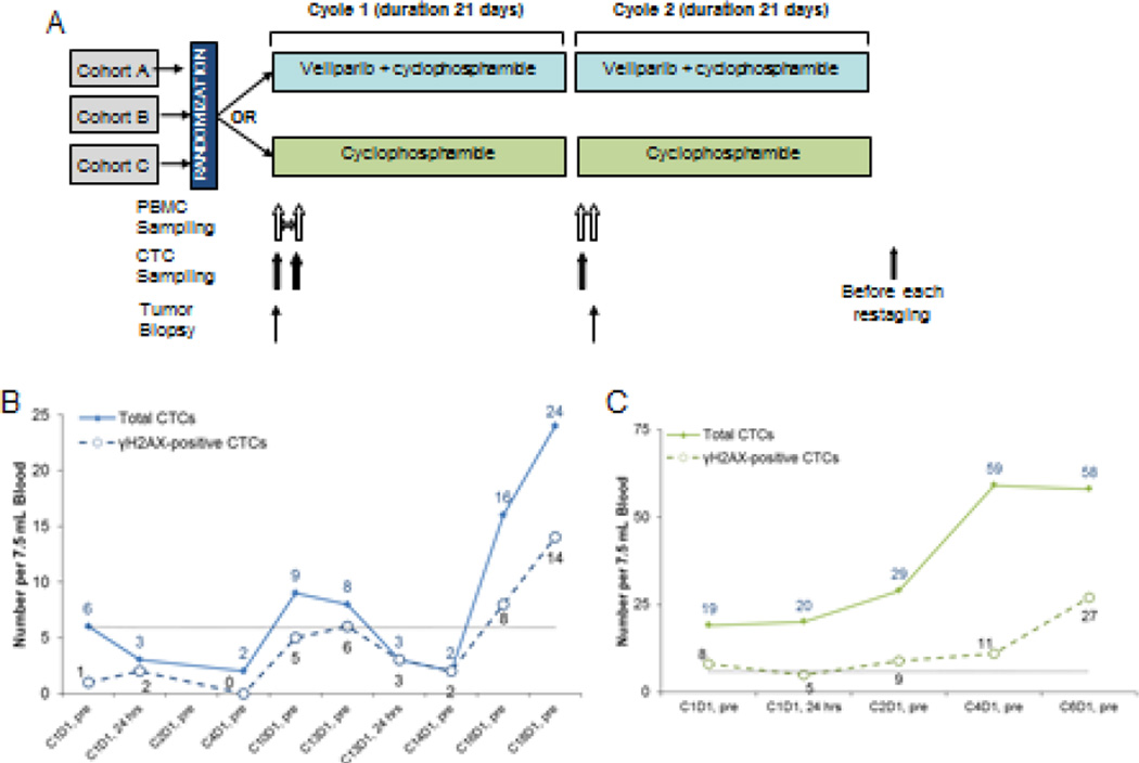
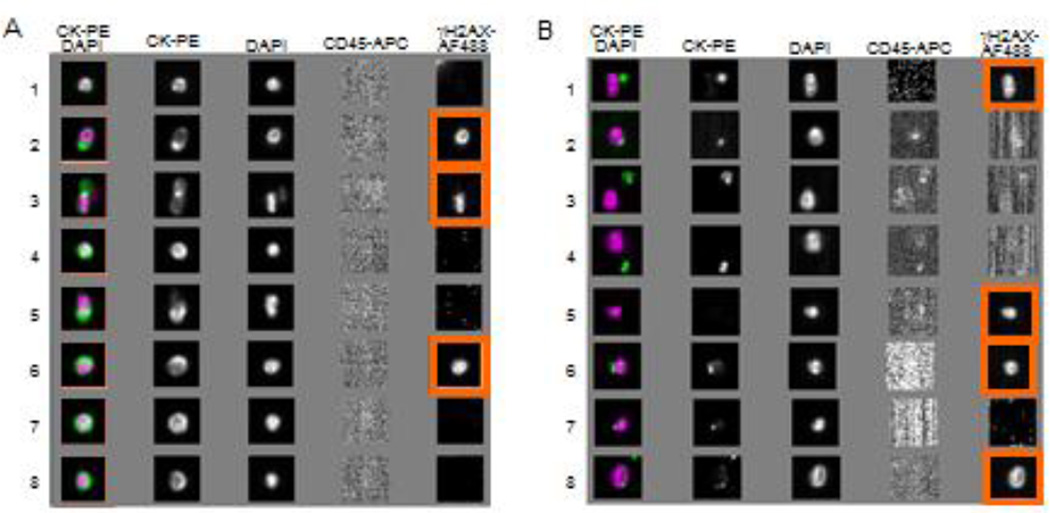
Circulating tumor cells (CTCs), which are captured from blood with anti-epithelial cell adhesion molecule (EpCAM) antibodies, have established prognostic value in specific epithelial cancers, but less is known about their utility for assessing patient response to molecularly targeted agents via measurement of pharmacodynamic (PD) endpoints. We discuss the use of CellSearch (Janssen Diagnostics, LLC, Raritan, NJ) CTC isolation technology for monitoring PD response in early phase trials. We present representative data from three clinical trials with the poly(ADP-ribose) polymerase (PARP) inhibitor veliparib (ABT-888) suggesting that CTCs can be used to measure PD effects. However, while often leading to hypothesis-generating information, our experience points to the difficulty in obtaining sufficient EpCAM-expressing CTCs from patients with advanced disease to reach statistically significant conclusions about PD effects from each trial. Overall, the level of phenotypic heterogeneity observed in specimens from patients with advanced carcinomas suggests caution in the use of cell-surface differentiation marker-based methods for isolating CTCs.

Keywords: Assay validation; CTCs; Pharmacodynamics; Surrogate endpoint.


Conflict of interest statement

None

Figures

Figure 1
Figure 1
Representative data on (A) total CTC count (normalized to 7.5 mL blood volume) and (B) proportion of γH2AX-positive CTCs enumerated from patients before and after treatment with topotecan (patient #1), topotecan and veliparib (patients #4, 5, and 6), or cyclophosphamide and veliparib (patients #2, 3, and 7–20). Grouped analysis indicated a significant difference in the proportion of γH2AX-positive CTCs before and 2 days after treatment (P = 0.0027). (C-D) Individual data for a patient with neuroendocrine prostate cancer treated with topotecan for 2 cycles. Adapted from Wang et al.
Figure 2
Figure 2
Baseline CTC counts in patients with advanced disease from the NCI Developmental Therapeutics Clinic (DTC) and other sites in the NCI clinical trials network. All patients from the NCI DTC had advanced refractory disease and were enrolled in the following clinical trials: NCT01306032, NCT01051635, NCT00978250, NCT01534598, NCT01748825, NCT01851369, NCT00923481, and NCT00900198. Patients from other sites were enrolled in NCT01264432, NCT01434316, NCT00034216, and NCT00576654. Whole blood samples (7.5 mL) were drawn from 381 patients with a variety of advanced malignancies enrolled in phase I or phase II clinical trials and CTC enumeration was performed by the CellSearch system. (A) Patients at the NCI DTC (N=238) had a mean of 29.1 (standard deviation, 144) and a median of 1.5 baseline CTC, while patients at other sites (N=143) had a mean of 162.5 (standard deviation, 1191) and median of 4 baseline CTCs per 7.5 mL blood. Median and interquartile range are plotted for each. (B) The frequencies of baseline CTC counts for patients at both the NCI DTC and other sites in the NCI clinical trials network (N=381) are shown grouped by cancer type. Median and interquartile range are plotted for each.
Figure 2
Figure 2
Baseline CTC counts in patients with advanced disease from the NCI Developmental Therapeutics Clinic (DTC) and other sites in the NCI clinical trials network. All patients from the NCI DTC had advanced refractory disease and were enrolled in the following clinical trials: NCT01306032, NCT01051635, NCT00978250, NCT01534598, NCT01748825, NCT01851369, NCT00923481, and NCT00900198. Patients from other sites were enrolled in NCT01264432, NCT01434316, NCT00034216, and NCT00576654. Whole blood samples (7.5 mL) were drawn from 381 patients with a variety of advanced malignancies enrolled in phase I or phase II clinical trials and CTC enumeration was performed by the CellSearch system. (A) Patients at the NCI DTC (N=238) had a mean of 29.1 (standard deviation, 144) and a median of 1.5 baseline CTC, while patients at other sites (N=143) had a mean of 162.5 (standard deviation, 1191) and median of 4 baseline CTCs per 7.5 mL blood. Median and interquartile range are plotted for each. (B) The frequencies of baseline CTC counts for patients at both the NCI DTC and other sites in the NCI clinical trials network (N=381) are shown grouped by cancer type. Median and interquartile range are plotted for each.
Figure 3
Figure 3
The percentage of baseline γH2AX-positive CTCs in patients with advanced disease from the NCI Developmental Therapeutics Clinic (DTC) and other sites in the NCI clinical trials network who exhibited 3 or more CTCs. Patient specimens with less than 3 baseline CTCs were not graphed because the fraction of γH2AX-positive CTCs in those samples is not a reliable measurement. All patients from the NCI DTC had advanced, refractory disease and were enrolled in trials NCT01306032, NCT01051635, NCT01748825, NCT01851369 and NCT00900198. Patients from other sites were enrolled in trials NCT01264432, NCT01434316, and NCT00576654. CTC enumeration by the CellSearch system on whole blood samples (7.5 mL) found 121 patients with a variety of advanced malignancies enrolled in phase I or phase II clinical trials. (A) At baseline, eligible patients at the NCI DTC (N=53) had, on average, 32% γH2AX-positive CTCs (standard deviation ±31%) and those from other sites (N=68) had, on average, 31% γH2AX-positive CTCs (standard deviation ±28%). Mean and standard deviation are plotted for each. (B) Baseline γH2AX-positive CTC frequencies grouped by cancer type. Mean and standard deviation are plotted for each.
Figure 4
Figure 4
Phase I Study of veliparib (ABT-888) in combination with LDFWAR therapy in patients with advanced solid malignancies with peritoneal carcinomatosis. (A) Schema of trial drug administration and 5 separate blood collection time points in cycle 1 of treatment used for CTC analysis. Blood for CTC analysis was collected at (1) C1D1, prior to LDFWAR, (2) C1D1, 6–8 hrs after LDFWAR, (3) C1D3, (4) C1D5, prior to veliparib and LDFWAR, and (5) C1D12 prior to veliparib and LDFWAR (B) CellSearch results for total CTC and γH2AX-positive CTCs over the course of therapy for two different patients. Black dotted line indicates recommended threshold of 6 CTCs for reporting biomarker-positive CTCs.
Figure 5
Figure 5
Phase I dose-escalation study of oral veliparib (ABT-888) plus intravenous irinotecan (CPT-11) administered in patients with advanced solid tumors. (A) Clinical trial drug administration schema indicating the 12 separate blood collection time points during treatment cycle 1 for CTC analysis. Blood for CTC analysis was collected at (1) screening; (2) C1D1 prior to irinotecan or veliparib; (3–5) 4–6 hrs, 8 hrs, and 22 hrs after single-agent irinotecan; and (6) C1D2, 24 hrs after irinotecan but before the first veliparib dose. After starting veliparib treatment, blood was also collected at (7–10) C1D8 prior to irinotecan, 4–6 hrs, 8 hrs, and 22 hrs after irinotecan; (11) C1D9, 24 hrs after irinotecan; and (12) C1D15. (B) Longitudinal tracking of total CTC numbers and proportion of γH2AX-positive CTCs in a treated patient. Blood from the C1D2 22 hr time point could not be analyzed due to hemolysis and clotting of the specimen (*). Black dotted line indicates recommended cut-off of 6 CTCs for reporting biomarker-positive CTCs.
Figure 6
Figure 6
Veliparib (ABT-888) with cyclophosphamide in refractory BRCA-positive ovarian, primary peritoneal or ovarian high-grade serous carcinoma, fallopian tube cancer, triple-negative breast cancer, and low-grade non-Hodgkin lymphoma. (A) Schema for clinical trial drug administration and CTC collection. (B-C) Total CTCs and γH2AX-positive CTCs for two patients initially on cyclophosphamide alone who then crossed over to the veliparib plus cyclophosphamide arm. Patient in panel B had breast cancer and crossed over to combination arm at cycle 13; patient in panel C had breast cancer and crossed over to combination arm at cycle 3. Both patients were taken off-study due to disease progression. Black dotted line indicates recommended cut-off of 6 CTCs for reporting biomarker-positive CTCs.
Figure 7
Figure 7
Images of CTCs and unassigned events by CellSearch. EpCAM antibody-enriched components from a blood specimen from a patient with advanced breast cancer were stained with DAPI and fluorescence antibodies CK-PE, CD45-APC, and γH2AX-AF488, and analyzed using the CellSearch system. For this specimen, 27 CTCs (EpCAM+/CK+/CD45/DAPI+) were detected and 7 of them were γH2AX-positive. Additionally, 906 unassigned events were observed in this specimen, among which 43 were CK/CD45/DAPI+ and, of those, 9 were γH2AX-positive. Images highlighted in orange show γH2AX-AF488 staining, indicating a γH2AX-positive CTC. (A) Images for 8 representative CTCs and (B) 8 unassigned events (not typical CTCs) are presented. Unassigned events (CK/CD45/DAPI+) were also captured by the EpCAM antibody in the specimen from the same patient.
Figure 8
Figure 8
CTCs detected from an ASPS patient blood sample using the CellSearch Circulating Endothelial Cell kit with anti-CD146 capture (and anti-CD105 phenotyping confirmation) on the CellSearch device. Staining with an antibody against the ASPL-TFE3 fusion protein in the last column authenticates the malignant origin of the cells.

References

    1. Allard WJ, Matera J, Miller MC, Repollet M, Connelly MC, Rao C, et al. Tumor cells circulate in the peripheral blood of all major carcinomas but not in healthy subjects or patients with nonmalignant diseases. Clin Cancer Res. 2004;10:6897–6904. - PubMed
    1. Cristofanilli M, Budd GT, Ellis MJ, Stopeck A, Matera J, Miller MC, et al. Circulating tumor cells, disease progression, and survival in metastatic breast cancer. N Engl J Med. 2004;351:781–791. - PubMed
    1. de Bono JS, Scher HI, Montgomery RB, Parker C, Miller MC, Tissing H, et al. Circulating tumor cells predict survival benefit from treatment in metastatic castration-resistant prostate cancer. Clin Cancer Res. 2008;14:6302–6309. - PubMed
    1. Cohen SJ, Punt CJ, Iannotti N, Saidman BH, Sabbath KD, Gabrail NY, et al. Relationship of circulating tumor cells to tumor response, progression-free survival, and overall survival in patients with metastatic colorectal cancer. J Clin Oncol. 2008;26:3213–3221. - PubMed
    1. Hayes DF, Cristofanilli M, Budd GT, Ellis MJ, Stopeck A, Miller MC, et al. Circulating tumor cells at each follow-up time point during therapy of metastatic breast cancer patients predict progression-free and overall survival. Clin Cancer Res. 2006;12:4218–4224. - PubMed

MeSH terms