. 2017 Jan;23(1):76-86.
doi: 10.1111/cns.12634. Epub 2016 Sep 24.

Levodopa Effect on Basal Ganglia Motor Circuit in Parkinson's Disease

Affiliations

Levodopa Effect on Basal Ganglia Motor Circuit in Parkinson's Disease

Lin-Lin Gao et al. CNS Neurosci Ther. 2017 Jan.

Abstract

Aims: To investigate the effects of levodopa on the basal ganglia motor circuit (BGMC) in Parkinson's disease (PD).

Methods: Thirty PD patients with asymmetrical bradykinesia and 30 control subjects were scanned using resting-state functional MRI. Functional connectivity of the BGMC was measured and compared before and after levodopa administration in patients with PD. The correlation between improvements in bradykinesia and changes in BGMC connectivity was examined.

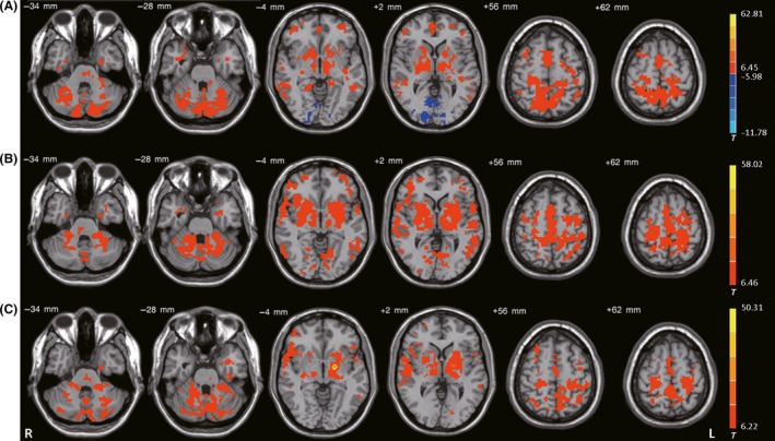
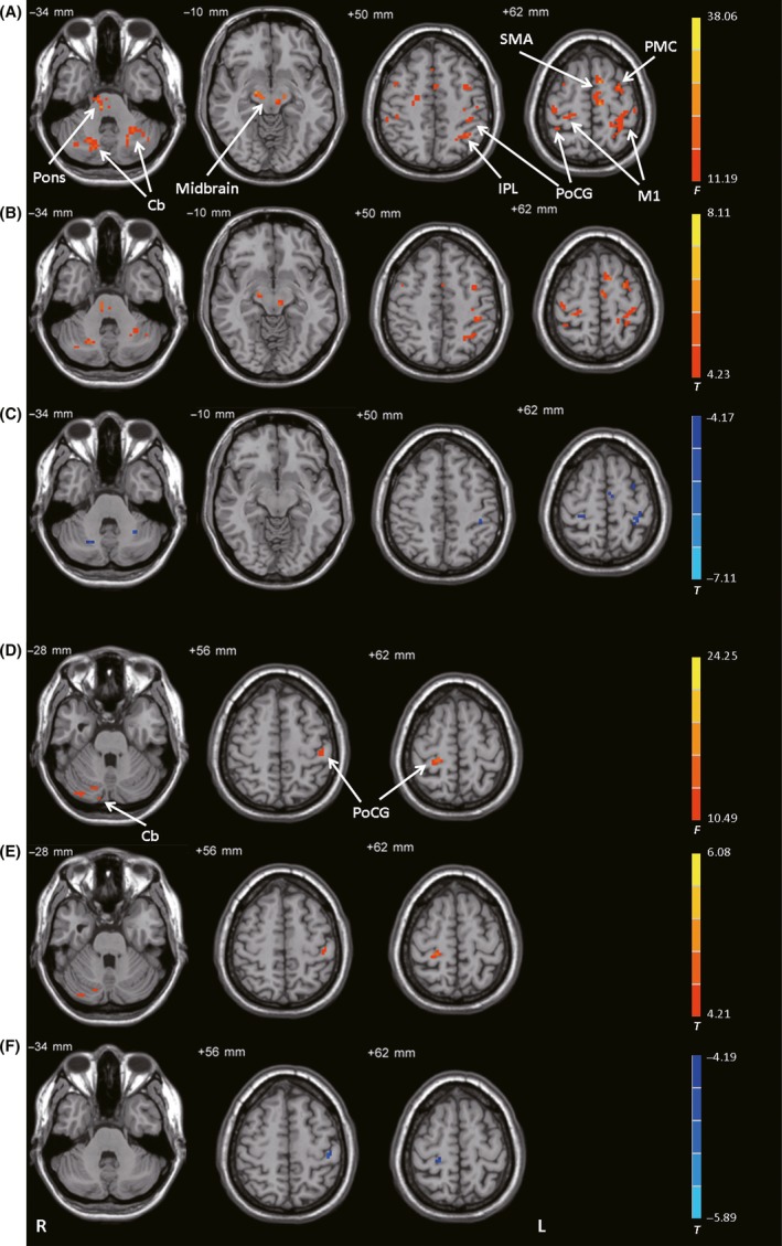
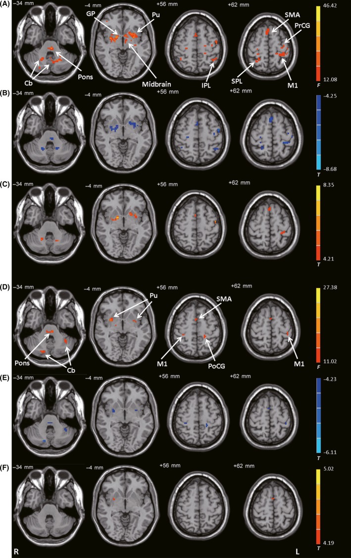
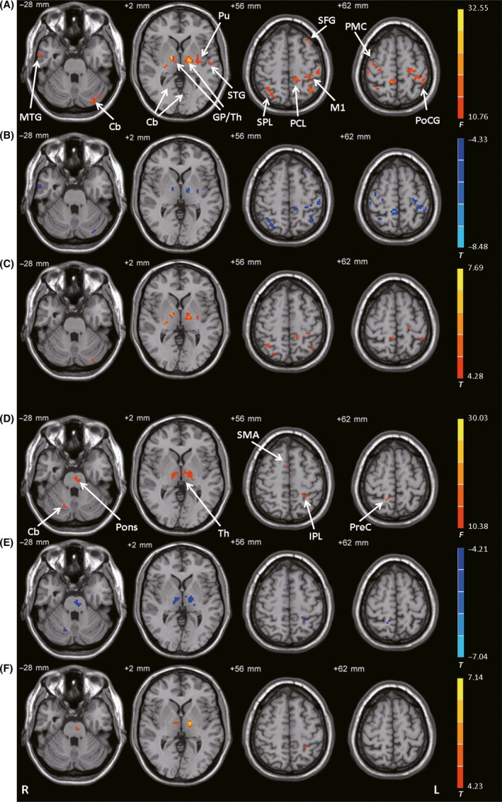
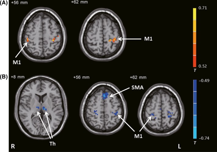
Results: In the PD-off state (before medication), the posterior putamen and internal globus pallidus (GPi) had decreased connectivity while the subthalamic nucleus (STN) had enhanced connectivity within the BGMC relative to control subjects. Levodopa administration increased the connectivity of posterior putamen- and GPi-related networks but decreased the connectivity of STN-related networks. Improvements in bradykinesia were correlated with enhanced connectivity of the posterior putamen-cortical motor pathway and with decreased connectivity of the STN-thalamo-cortical motor pathway.

Conclusion: In PD patients with asymmetrical bradykinesia, levodopa can partially normalize the connectivity of the BGMC with a larger effect on the more severely affected side. Moreover, the beneficial effect of levodopa on bradykinesia is associated with normalization of the striato-thalamo-cortical motor and STN-cortical motor pathways. Our findings inform the neural mechanism of levodopa treatment in PD.

Keywords: Basal ganglia motor circuit; Bradykinesia; Connectivity; Levodopa; Parkinson's disease.


Conflict of interest statement

The authors have no conflict of interests to disclose.

Figures

Figure 1
Figure 1
Functional connectivity among the 3 ROIs. Brain regions showing significant connectivity with the (A) left subthalamic nucleus, (B) left posterior putamen, and (C) left internal globus pallidus in healthy control subjects (one‐sample t‐test, P < 1 × 10⁻⁵, FDR‐corrected). T‐value bar is shown on the right. Warm and cold tones indicate positive and negative connectivity with the ROIs, respectively. L, left; R, right.
Figure 2
Figure 2
Connectivity with the STN. (A) Brain regions showing significant differences in connectivity with the left STN among control subjects, patients with PD before levodopa administration (PD‐off), and patients with PD after levodopa administration (PD‐on) (ANCOVA, P < 0.05, FDR‐corrected). (B) Increased connectivity with the left STN in the PD‐off condition versus the control condition (post hoc analysis, P < 0.05, FDR‐corrected). (C) Decreased connectivity with the left STN in the PD‐on condition versus the PD‐off condition (post hoc analysis, P < 0.05, FDR‐corrected). (D) Brain regions showing significant differences in connectivity with the right STN among the control, PD‐off, and PD‐on conditions (ANCOVA, P < 0.05, FDR‐corrected). (E) Increased connectivity with the right STN in the PD‐off condition versus the control condition (post hoc analysis, P < 0.05, FDR‐corrected). (F) Decreased connectivity with the right STN in the PD‐on condition versus the PD‐off condition (post hoc analysis, P < 0.05, FDR‐corrected). The warm and cold tones indicate increases and decreases in connectivity, respectively. F‐ or T‐value bars are shown on the right. Cb, cerebellum; IPL, inferior parietal lobule; L, left; M1, primary motor cortex; PMC, premotor cortex; PoCG, postcentral gyrus; R, right; SMA, supplementary motor area.
Figure 3
Figure 3
Connectivity with the posterior putamen. (A) Brain regions showing significant differences in connectivity with the left posterior putamen among the control, PD‐off, and PD‐on conditions (ANCOVA, P < 0.05, FDR‐corrected). (B) Decreased connectivity with the left posterior putamen in the PD‐off condition versus the control condition (post hoc analysis, P < 0.05, FDR‐corrected). (C) Increased connectivity with the left posterior putamen in the PD‐on condition versus the PD‐off condition (post hoc analysis, P < 0.05, FDR‐corrected). (D) Brain regions showing significant differences in connectivity with the right posterior putamen among the control, PD‐off, and PD‐on conditions (ANCOVA, P < 0.05, FDR‐corrected). (E) Decreased connectivity with the right posterior putamen in the PD‐off condition versus the control condition (post hoc analysis, P < 0.05, FDR‐corrected). (F) Increased connectivity with the right posterior putamen in the PD‐on condition versus the PD‐off condition (post hoc analysis, P < 0.05, FDR‐corrected). F‐ or T‐ value bars are shown on the right. Cb, cerebellum; GP, globus pallidus; IPL, inferior parietal lobule; L, left; M1, primary motor cortex; PoCG, postcentral gyrus; PrCG, precentral gyrus; Pu, putamen; R, right; SMA, supplementary motor area; SPL, superior parietal lobule.
Figure 4
Figure 4
Connectivity with the GPi. (A) Brain regions showing significant differences in connectivity with the left GPi among the control, PD‐off, and PD‐on conditions (ANCOVA, P < 0.05, FDR‐corrected). (B) Decreased connectivity with the left GPi in the PD‐off condition versus the control condition (post hoc analysis, P < 0.05, FDR‐corrected). (C) Increased connectivity with the left GPi in the PD‐on condition versus the PD‐off condition (post hoc analysis, P < 0.05, FDR‐corrected). (D) Brain regions showing significant differences in connectivity with the right GPi among the control, PD‐off, and PD‐on conditions (ANCOVA, P < 0.05, FDR‐corrected). (E) Decreased connectivity with the right GPi in the PD‐off condition versus the control condition (post hoc analysis, P < 0.05, FDR‐corrected). (F) Increased connectivity with the right GPi in the PD‐on condition versus the PD‐off condition (post hoc analysis, P < 0.05, FDR‐corrected). F‐ or T‐value bars are shown on the right. Cb, cerebellum; GP, globus pallidus; IPL, inferior parietal lobule; L, left; M1, primary motor cortex; MTG, middle temporal gyrus; PCL, paracentral lobule; PMC, premotor cortex; PoCG, postcentral gyrus; PreC, precuneus; Pu, putamen; R, right; SMA, supplementary motor area; SPL, superior parietal lobule; STG, superior temporal gyrus; Th, thalamus.
Figure 5
Figure 5
Results of the correlation analysis. Brain regions showing significant correlations with bradykinesia subscale score between the PD‐on and PD‐off conditions against differences in connectivity between the PD‐on and PD‐off conditions (ΔFC) in the left posterior putamen (A) and left STN (B) (correlation analysis, P < 0.05, FDR‐corrected). The warm and cold tones indicate positive and negative correlations, respectively. T‐value bar is shown on the right. L, left; M1, primary motor cortex; R, right; SMA, supplementary motor area; Th, thalamus.

Similar articles

Cited by

References

    1. Hauser RA. Levodopa: Past, present, and future. Eur Neurol 2009;62:1–8. - PubMed
    1. Haslinger B, Erhard P, Kampfe N, et al. Event‐related functional magnetic resonance imaging in Parkinson's disease before and after levodopa. Brain 2001;124:558–570. - PubMed
    1. Wu T, Hallett M. A functional MRI study of automatic movements in patients with Parkinson's disease. Brain 2005;128:2250–2259. - PubMed
    1. Herz DM, Eickhoff SB, Lokkegaard A, Siebner HR. Functional neuroimaging of motor control in Parkinson's disease: A meta‐analysis. Hum Brain Mapp 2014;35:3227–3237. - PMC - PubMed
    1. Wu T, Wang L, Hallett M, Chen Y, Li K, Chan P. Effective connectivity of brain networks during self‐initiated movement in Parkinson's disease. NeuroImage 2011;55:204–215. - PubMed

MeSH terms