Review
. 2017 Jan;112(Pt A):11-15.
doi: 10.1016/j.neuropharm.2016.08.034. Epub 2016 Sep 20.

Structure and symmetry inform gating principles of ionotropic glutamate receptors

Affiliations
Review

Structure and symmetry inform gating principles of ionotropic glutamate receptors

Shujia Zhu et al. Neuropharmacology. 2017 Jan.

Abstract

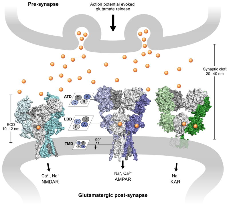
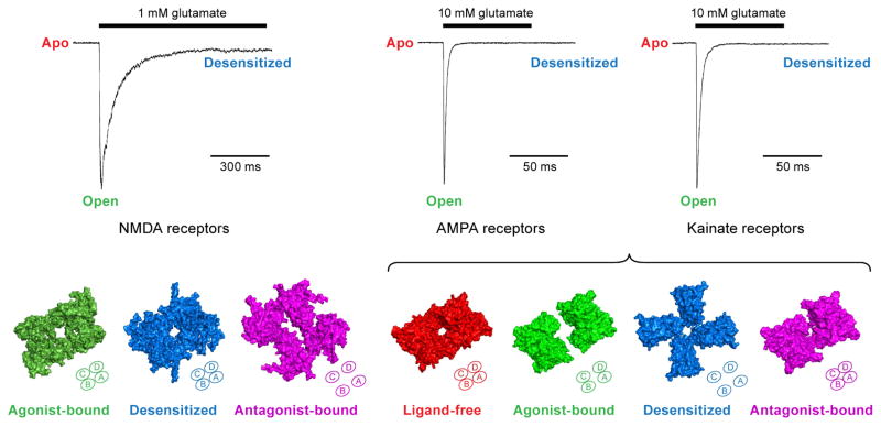
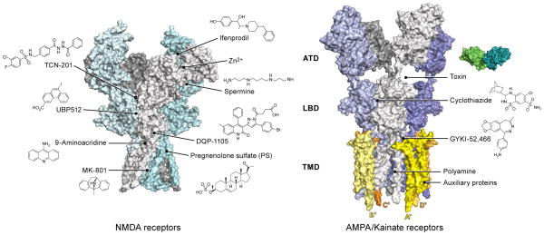
Ionotropic glutamate receptors (iGluRs) transduce signals derived from release of the excitatory neurotransmitter glutamate from pre-synaptic neurons into excitation of post-synaptic neurons on a millisecond time-scale. In recent years, the elucidation of full-length iGluR structures of NMDA, AMPA and kainate receptors by X-ray crystallography and single particle cryo-electron microscopy has greatly enhanced our understanding of the interrelationships between receptor architecture and gating mechanism. Here we briefly review full-length iGluR structures and discuss the similarities and differences between NMDA receptors and non-NMDA iGluRs. We focus on distinct conformations, including ligand-free, agonist-bound active, agonist-bound desensitized and antagonist-bound conformations as well as modulator and auxiliary protein-bound states. These findings provide insights into structure-based mechanisms of iGluR gating and modulation which together shape the amplitude and time course of the excitatory postsynaptic potential. This article is part of the Special Issue entitled 'Ionotropic glutamate receptors'.


Figures

Figure 1
Figure 1. iGluRs at glutamatergic synapses
The action potential evokes release of the excitatory neurotransmitter glutamate (shown as yellow cycles) from pre-synaptic vesicles. Structures of three main iGluRs (surface representation) are shown in the post-synaptic membrane. From left to right: NMDA receptor in the glycine/glutamate bound state (PDB: 5IOU), AMPA receptor in the competitive antagonist MPQX bound state (PDB: 5KK2) and kainate receptor in the presence of the agonist 2S,4R-4-methylglutamate (PDB: 4UQQ). iGluRs are heteromeric (NMDA receptors) and/or homomeric (AMPA/kainate receptors) dimer-of-dimers assemblies, with each subunit arranged in three layers defined by the amino-terminal domain (ATD), the ligand-binding domain (LBD) and the transmembrane domain (TMD). The subunit cross-talk on the ATD and LBD layers is illustrated in the cartoon on the left. The lengths of the synaptic cleft and the extracelluar domain (ECD) of the iGluRs are also marked.
Figure 2
Figure 2. Conformational changes of the LBD gating ‘ring’ during gating cycle
Top panel: Patch-clamp recordings of iGluR current, where NMDA receptors show slow desensitization kinetics and AMPA/kainate receptors display fast gating kinetics. Bottom panel: Top-down views of different ligand-binding domain (LBD) structures in physiologically-relevant conformations that correspond to the agonist-bound desensitized state (PDB: 5IOU) and antagonist-bound closed state (PDB: 5IPS) of the NMDA receptor; the ligand-free state (PDB: 4U2P), agonist-bound open state (PDB: 4UQK), antagonist-bound closed state (PDB: 3KG2) of the AMPA receptor; and the agonist-bound desensitized state (PDB: 4UQQ) of the kainate receptor. The tetrameric arrangement of the LBD gating ring in each state is also illustrated in diagrams at the bottom right corner of each structure.
Figure 3
Figure 3. Pharmacological properties and auxiliary proteins
iGluRs harbor multiple binding sites for small molecules acting as allosteric modulators and for auxiliary proteins that modulate receptor trafficking and activity. The left panel shows how NMDA receptors have binding pockets on ATD, LBD and TMD layers. The right panel shows that AMPA/kainate receptors have binding cavities between the ATD-LBD layer, within the LBD dimer interface, and within the TMDs for both small molecules and proteins.

References

    1. Acker TM, Yuan H, Hansen KB, Vance KM, Ogden KK, Jensen HS, Burger PB, Mullasseri P, Synder JP, Liotta DC, Traynelis SF. Mechanism for noncompetitive inhibition by novel GluN2C/D N-methyl-D-asparate receptor subunit-selective modulators. Mol Pharmacol. 2011;80:782–795. - PMC - PubMed
    1. Attwell D, Gibb A. Neuroenergetics and the kinetic design of excitatory synapses. Nature Rev Neurosci. 2005;6:841–849. - PubMed
    1. Chen L, Dürr K, Gouaux E. X-ray structures of AMPA receptor-cone snail toxin complexes illuminate activation mechanisms. Science. 2014;345:1021–1026. - PMC - PubMed
    1. Dürr K, Chen L, Stein RA, De Zorzi R, Folea IM, Walz T, Mchaourab HS, Gouaux E. Structure and dynamics of AMPA receptor GluA2 in resting, pre-open and desensitized states. Cell. 2014;158:778–792. - PMC - PubMed
    1. Hackos DH, Hanson JE. Diverse modes of NMDA receptor positive allosteric modulatioin: Mechanisms and consequences. Neuropharmacology. 2016 doi:10.1016j. - PubMed

MeSH terms

Substances