. 2016 Nov 1;311(5):H1234-H1247.
doi: 10.1152/ajpheart.00235.2016. Epub 2016 Sep 23.

The chromatin-binding protein Smyd1 restricts adult mammalian heart growth

Affiliations

The chromatin-binding protein Smyd1 restricts adult mammalian heart growth

Sarah Franklin et al. Am J Physiol Heart Circ Physiol. .

Abstract

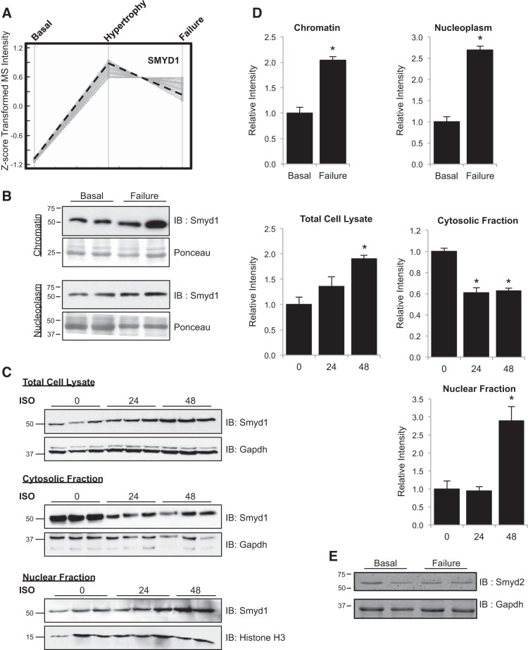
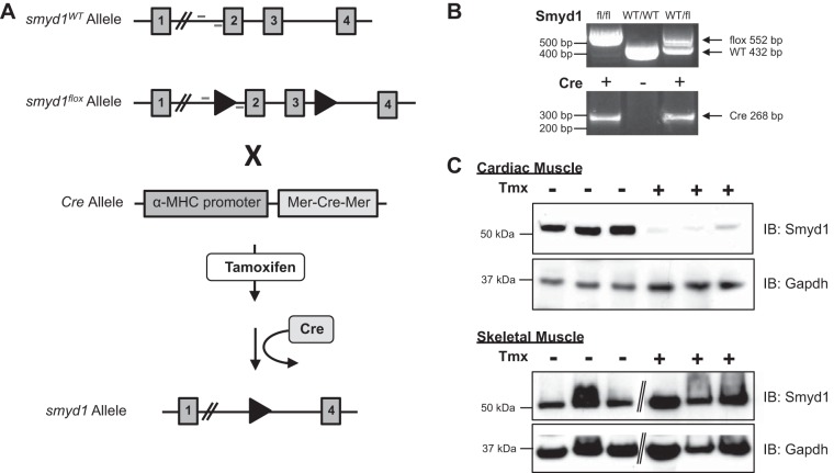
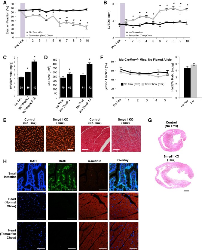
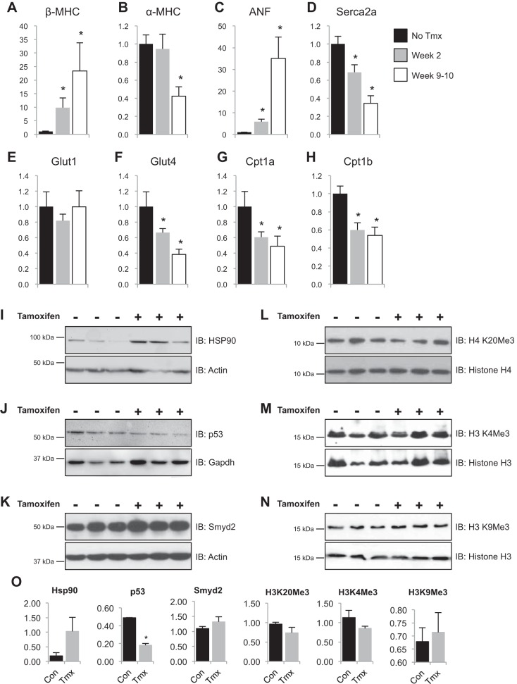
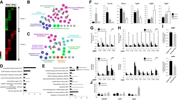
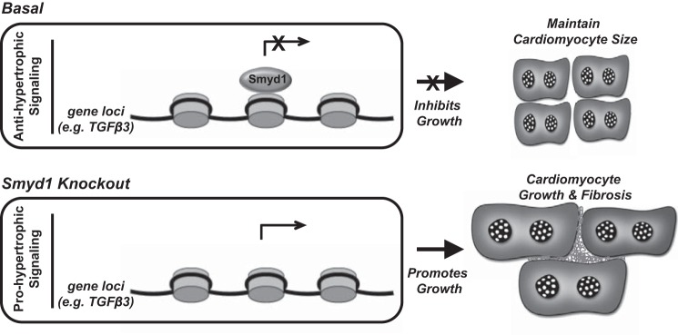
All terminally differentiated organs face two challenges, maintaining their cellular identity and restricting organ size. The molecular mechanisms responsible for these decisions are of critical importance to organismal development, and perturbations in their normal balance can lead to disease. A hallmark of heart failure, a condition affecting millions of people worldwide, is hypertrophic growth of cardiomyocytes. The various forms of heart failure in human and animal models share conserved transcriptome remodeling events that lead to expression of genes normally silenced in the healthy adult heart. However, the chromatin remodeling events that maintain cell and organ size are incompletely understood; insights into these mechanisms could provide new targets for heart failure therapy. Using a quantitative proteomics approach to identify muscle-specific chromatin regulators in a mouse model of hypertrophy and heart failure, we identified upregulation of the histone methyltransferase Smyd1 during disease. Inducible loss-of-function studies in vivo demonstrate that Smyd1 is responsible for restricting growth in the adult heart, with its absence leading to cellular hypertrophy, organ remodeling, and fulminate heart failure. Molecular studies reveal Smyd1 to be a muscle-specific regulator of gene expression and indicate that Smyd1 modulates expression of gene isoforms whose expression is associated with cardiac pathology. Importantly, activation of Smyd1 can prevent pathological cell growth. These findings have basic implications for our understanding of cardiac pathologies and open new avenues to the treatment of cardiac hypertrophy and failure by modulating Smyd1.

Keywords: Smyd1; cardiac hypertrophy; epigenetics; heart failure; histone methyltransferase.


Figures

Fig. 1.
Fig. 1.
Identification of Smyd1 as a novel participant in pathological cardiac hypertrophy and failure. A: among hundreds of chromatin-bound proteins quantified during pressure overload-induced hypertrophy and heart failure, Smyd1 abundance increased during compensatory hypertrophy and failure, as measured by mass spectrometry-based peptide abundance from cardiac chromatin. This trend was confirmed by Western blotting (B); we also observed an increase in nucleoplasmic (i.e., nuclear localized, not bound to chromatin) Smyd1 during heart failure. IB, immunoblot. C: to determine whether activation of Smyd1 was conserved across species and stimulus, we examined expression and localization following isoproterenol (ISO; 1 μM for times indicated) treatment of ventricular myocytes. D: quantitation of Western blots from B and C. E: Smyd2 expression is unchanged in the setting of pressure overload-induced heart failure; n = 4–6 per group for all Westerns; bars are SE, *P < 0.05.
Fig. 2.
Fig. 2.
Generation of cardiac-specific, tamoxifen-inducible smyd1-null mice. A: positional cloning was used to insert loxp sites flanking exons 2 and 3 of smyd1 to produce the target allele. These mice were bred to homozygosity and then crossed with transgenic mice expressing the Cre recombinase positioned between two copies of the tamoxifen-responsive modified estrogen receptor and driven by the α-myosin heavy chain (MHC) promoter (so called Mer-Cre-Mer mice). B: PCR genotyping confirmed genetic manipulation (location of primers are indicated in A). Resulting mice express Mer-Cre-Mer in adult cardiomyocytes, and, upon treatment with tamoxifen (Tmx), this protein excises the floxed region of the mutant smyd1 allele. Mice fed a tamoxifen diet for 3 wk followed by 1 wk on a normal diet demonstrate near complete loss of Smyd1 protein specifically in the heart; Smyd1 in skeletal muscle is unaffected (C). Diagonal lines indicate removal of gel lanes. WT, wild-type.
Fig. 3.
Fig. 3.
Loss of Smyd1 in the adult heart induces progressive hypertrophy and failure at the organ and cell level. Cardiac function in smyd1flox/floxCre+/− mice was measured by echocardiography (ECHO). A baseline reading was taken in each mouse before 3 wk on tamoxifen or regular diet. After 1 wk back on regular diet in the experimental group, ECHO measurements were made on a weekly basis in both groups. We observed a progressive decline in left ventricular ejection fraction (A) coupled with a progressive increase in left ventricular end-diastolic dimension (LVEDd) (B) as a result of smyd1 deletion (n = 37 control mice and 36 tamoxifen=treated mice for A and B; *P < 0.05 vs. regular diet at same time point). Loss of Smyd1 also induced an increase in muscle mass at the whole organ (as measured by heart weight:body weight ratio, HW/BW, *P < 1E-5; C) and individual cardiac cell level as measured following wheat germ agglutinin staining (D and E, left, 10 wk after tmx; bar = 50 μm), concomitant with an increase in fibrotic deposition as measured by trichrome staining (E, right, 8 wk after tmx; bar = 100 μm); n values for HW/BW and cell size are indicated, the former indicating number of animals and the latter indicating number of cells (from a total of 4 animals per group). KO, knockout. F: MerCreMer-smyd1wt/wt mice (i.e., mice with no floxed alleles) develop neither cardiac dysfunction nor hypertrophy when administered the same tamoxifen protocol. G: marked chamber dilation is observed in Smyd1-deficient mice at 8–10 wk after return to normal chow. H: MerCreMer-smyd1flox/flox mice were injected with 5-bromo-1-(2-deoxy-β-d-ribofuranosyl) (BrdU) to label actively replicating cells. Small intestine positive control shows strong BrdU incorporation. We observe incorporation of BrdU in neither the hearts of MerCreMer-smyd1flox/flox mice fed a normal diet (middle) nor those with the same genotype on a tamoxifen diet for 10 wk (bottom; bar = 50 μm).
Fig. 4.
Fig. 4.
Smyd1 is a previously unknown regulator of a conserved transcriptional program underlying heart failure. AH: fetal genes were measured by RT-PCR and values expressed as fold change relative to mice not fed tamoxifen. *P < 0.05; n = 5–8 for no tmx, n = 6 for post tmx 2 wk, and n = 7 for post tmx 9–10 wk. ANF, atrial natriuretic factor. IN: as determined by Western blotting, loss of Smyd1 increased expression of Hsp90, decreased expression of p53, resulted in an upregulation of Smyd2, and had no effect on global levels of histone H4 (K20) and H3 (K4 and K9) trimethylation. O: quantitation of IN; *P = 0.002.
Fig. 5.
Fig. 5.
Transcriptome analyses reveal Smyd1 to be a transcriptional repressor of a core set of developmental genes. Transcriptome analyses were performed separately on right ventricles (RV) and left ventricles (LV) from mice with deletion of Smyd1 at 2 wk and 9 wk after removal from tamoxifen diet (controls were normal diet-fed littermates). A: all genes measured in both groups, RV and LV, are displayed in heat map format (red is upregulation, green downregulation, and black statistically unchanged) and clustered according to similar behavior. 2 clusters were defined as indicated, and the functionality of genes in those clusters was determined by gene ontology (GO) analysis. The networks for cluster 1 (B) and 2 (C) for the biological process ontology are shown. In the network figures, node size indicates the P value, and linkage between two terms indicates the relatedness, as determined by κ statistics. The color indicates functional groups with each group represented by their most significant leading term. All terms shown in the network image of cluster 1 are filtered at P < 0.005; those in cluster 2 are filtered at P < 0.0001. Bioinformatic analyses of transcripts with altered expression in the LV, at week 2 (D) and week 9 (E) after Tmx treatment, show significant enrichment in genes involved in extracellular matrix remodeling, fibrosis, and transcriptional repression. MF, molecular function; BP = biological process; CC, cellular component; KEGG, KEGG analysis; ITP, Interpro analysis. Microarray results for several of these transcripts were subsequently validated by RT-PCR (F: all those we attempted to validate are shown; n = 3–6/group; *P < 0.05 vs no tamoxifen group). Chromatin immunoprecipitation (ChIP) and qPCR for Smyd1 (using FLAG antibody) show enrichment in the promoter region of target genes tgfbeta3 (G) and nppa (H); however, no corresponding enrichment of histone H3 lysine K4 trimethylation was detected in these regions (bottom). *P ≤ 0.05. I: luciferase reporter assay using the tgfbeta3 and nppa promoters confirms that Smyd1 acts as a transcriptional repressor by inhibiting transcription of these genes; n = 6/group; *P < 0.05. J: as a negative control, Smyd1 is enriched by ChIP-PCR at neither β-tubulin nor β-actin using primers shown previously to target the regulatory regions upstream of these genes [−3 kb for β-tubulin (24), −73 bp for β-actin (29)].
Fig. 6.
Fig. 6.
Overexpression of Smyd1 attenuates cellular hypertrophy in vivo. A: there are 2 splice variants of Smyd1 in mouse heart, differing by a 13-amino-acid sequence present in Smyd1a and absent in Smyd1b, but conserved in the single human Smyd1 transcript. BD: adenoviral infection of myocytes led to robust Smyd1 expression, as confirmed by RT-PCR and Western blotting. MOI, multiplicity of infection. E and F: overexpression of Smyd1a had no effect on basal cell size but prevented phenylephrine (PE)-induced myocyte hypertrophy (phalloidin staining; scale bar = 100 μm; quantified from ∼100 cells per group in 3 independent experiments; *P < 0.001). G and H: overexpression of Smyd1b required higher MOI and had no effect on PE-induced cell growth (quantified as described in F). I: RT-PCR analysis of fetal genes and Smyd1 targets in neonatal rat ventricular myocytes after adenovirus infection in the presence and absence of PE. *P ≤ 0.05 compared with empty virus control without PE.
Fig. 7.
Fig. 7.
Model for functions of Smyd1 in the adult myocardium. Under basal conditions, Smyd1 functions as a transcriptional repressor to inhibit cell growth and maintain cardiomyocyte size. Loss of Smyd1 in the adult myocardium leads to prohypertrophic signaling resulting in myocyte growth, fibrosis, and functional decline.

References

    1. Abaci N, Gulec C, Bayrak F, Komurcu Bayrak E, Kahveci G, Erginel Unaltuna N. The variations of BOP gene in hypertrophic cardiomyopathy. Anadolu Kardiyol Derg 10: 303–309, 2010. - PubMed
    1. Abu-Farha M, Lambert JP, Al-Madhoun AS, Elisma F, Skerjanc IS, Figeys D. The tale of two domains: Proteomics and genomics analysis of SMYD2, a new histone methyltransferase. Mol Cell Proteomics 7: 560–572, 2008. - PubMed
    1. Ago T, Liu T, Zhai P, Chen W, Li H, Molkentin JD, Vatner SF, Sadoshima J. A redox-dependent pathway for regulating class II HDACs and cardiac hypertrophy. Cell 133: 978–993, 2008. - PubMed
    1. Anand P, Brown JD, Lin CY, Qi J, Zhang R, Artero PC, Alaiti MA, Bullard J, Alazem K, Margulies KB, Cappola TP, Lemieux M, Plutzky J, Bradner JE, Haldar SM. BET bromodomains mediate transcriptional pause release in heart failure. Cell 154: 569–582, 2013. - PMC - PubMed
    1. Bindea G, Mlecnik B, Hackl H, Charoentong P, Tosolini M, Kirilovsky A, Fridman WH, Pages F, Trajanoski Z, Galon J. ClueGO: A Cytoscape plug-in to decipher functionally grouped gene ontology and pathway annotation networks. Bioinformatics 25: 1091–1093, 2009. - PMC - PubMed

Publication types

MeSH terms