. 2016 Sep 23;18(1):95.
doi: 10.1186/s13058-016-0749-6.

PRKCQ promotes oncogenic growth and anoikis resistance of a subset of triple-negative breast cancer cells

Affiliations

PRKCQ promotes oncogenic growth and anoikis resistance of a subset of triple-negative breast cancer cells

Jessica Byerly et al. Breast Cancer Res. .

Abstract

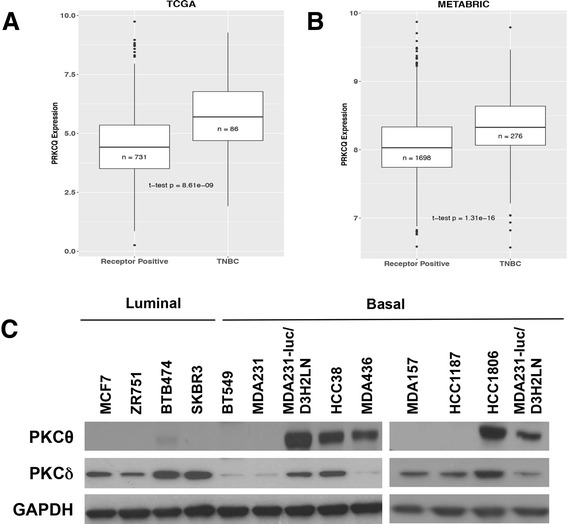
Background: The protein kinase C (PKC) family comprises distinct classes of proteins, many of which are implicated in diverse cellular functions. Protein tyrosine kinase C theta isoform (PRKCQ)/PKCθ, a member of the novel PKC family, may have a distinct isoform-specific role in breast cancer. PKCθ is preferentially expressed in triple-negative breast cancer (TNBC) compared to other breast tumor subtypes. We hypothesized that PRKCQ/PKCθ critically regulates growth and survival of a subset of TNBC cells.

Methods: To elucidate the role of PRKCQ/PKCθ in regulating growth and anoikis resistance, we used both gain and loss of function to modulate expression of PRKCQ. We enhanced expression of PKCθ (kinase-active or inactive) in non-transformed breast epithelial cells (MCF-10A) and assessed effects on epidermal growth factor (EGF)-independent growth, anoikis, and migration. We downregulated expression of PKCθ in TNBC cells, and determined effects on in vitro and in vivo growth and survival. TNBC cells were also treated with a small molecule inhibitor to assess requirement for PKCθ kinase activity in the growth of TNBC cells.

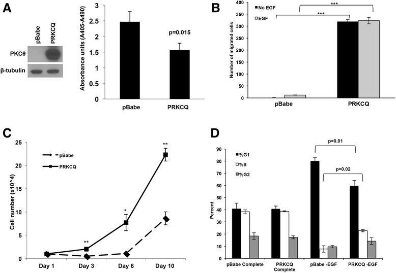
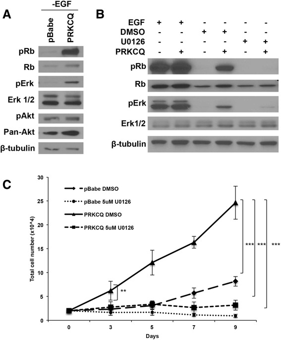
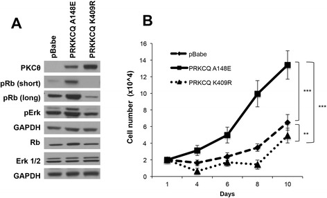
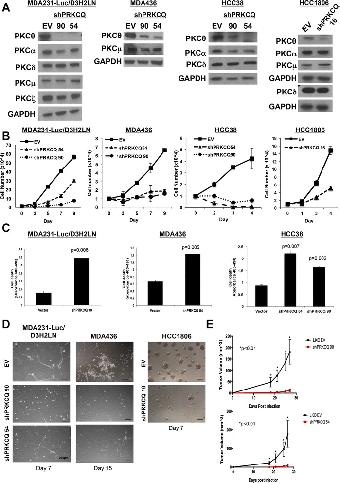
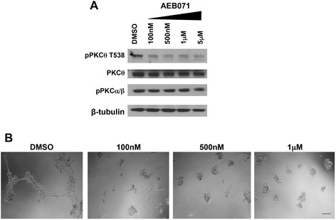
Results: PRKCQ/PKCθ can promote oncogenic phenotypes when expressed in non-transformed MCF-10A mammary epithelial cells; PRKCQ/PKCθ enhances anchorage-independent survival, growth-factor-independent proliferation, and migration. PKCθ expression promotes retinoblastoma (Rb) phosphorylation and cell-cycle progression under growth factor-deprived conditions that typically induce cell-cycle arrest of MCF-10A breast epithelial cells. Proliferation and Rb phosphorylation are dependent on PKCθ-stimulated extracellular signal-related kinase (Erk)/mitogen-activated protein kinase (MAPK) activity. Enhanced Erk/MAPK activity is dependent on the kinase activity of PKCθ, as overexpression of kinase-inactive PKCθ does not stimulate Erk/MAPK or Rb phosphorylation or promote growth-factor-independent proliferation. Downregulation of PRKCQ/PKCθ in TNBC cells enhances anoikis, inhibits growth in 3-D MatrigelTM cultures, and impairs triple-negative tumor xenograft growth. AEB071, an inhibitor of PKCθ kinase activity, also inhibits growth and invasive branching of TNBC cells in 3-D cultures, further supporting a role for PKCθ kinase activity in triple-negative cancer cell growth.

Conclusions: Enhanced PRKCQ/PKCθ expression can promote growth-factor-independent growth, anoikis resistance, and migration. PRKCQ critically regulates growth and survival of a subset of TNBC. Inhibition of PKCθ kinase activity may be an attractive therapeutic approach for TNBC, a subtype in need of improved targeted therapies.

Keywords: Anoikis; EGF-independent growth; PRKCQ/PKCθ; Triple-negative breast cancer.


Figures

Fig. 1
Fig. 1
Protein tyrosine kinase C theta isoform (PRKCQ) overexpression promotes multiple oncogenic activities in breast epithelial cells. a Non-transformed, immortalized MCF-10A cells overexpressing vector control or PRKCQ were generated, and assessed for their ability to survive in suspension cultures using the Cell Death ELISA assay. b The ability of MCF-10A cells overexpressing vector control or PRKCQ in Transwell Boyden chamber assays in the presence or absence of epidermal growth factor (EGF) was evaluated. The number of cells that migrated 18 hours after plating in the insert was counted. Five fields of migrated cells (×10 objective) were counted and averaged. c MCF-10A cells expressing vector control or PRKCQ (1 × 10^4 cells) were plated on day 1 in growth media lacking EGF. The number of cells were counted on the indicated days and plotted. d Cell-cycle profile analysis of empty vector or PRKCQ-expressing cells was performed by fluorescence-activated cell sorting after propidium iodide staining of cells cultured in growth media +/- EGF: *p < 0.05, **p < 0.01, ***p < 0.001. PKC protein kinase C
Fig. 2
Fig. 2
Extracellular signal-related (Erk)/mitogen-activated protein kinase (MAPK) activation is required for protein tyrosine kinase C theta isoform (PRKCQ)-mediated epidermal growth factor (EGF)-independent growth. a MCF-10A cells overexpressing pBabe vector control or wild-type PRKCQ were cultured in growth media lacking EGF. Lysates were probed with the indicated antibodies. b PRKCQ or vector control expressing MCF-10A cells were cultured in growth media +/- EGF. Cells grown in the absence of EGF were treated for 24 hours with dimethyl sulfoxide (DMSO) or mitogen-activated protein kinase kinase (MEK) inhibitor U0126 (5 μM). Cells were lysed and probed with the indicated antibodies. c MCF-10A cells expressing vector control or PRKCQ were cultured in growth media lacking EGF and treated with DMSO or U0126 (5 μM) for the indicated number of days. DMSO or U0126 was replaced every 2–3 days. Cell numbers were counted on the indicated days and plotted. Rb retinoblastoma
Fig. 3
Fig. 3
Protein tyrosine kinase C theta isoform (PRKCQ) kinase activity is required for epidermal growth factor (EGF)-independent growth, Extracellular signal-related kinase (Erk activity) and retinoblastoma (Rb) phosphorylation. a MCF-10A cells expressing vector control, constitutively active (A148E) or kinase-inactive (K409R) PKCθ, were cultured in growth media lacking EGF. Cell lysates were probed with the indicated antibodies. b MCF-10A cells expressing vector control, active or kinase-dead PKCθ, were cultured in media lacking EGF for the indicated number of days and counted
Fig. 4
Fig. 4
Protein tyrosine kinase C theta isoform (PRKCQ) is preferentially expressed in the triple-negative breast cancer (TNBC) subtype and breast cancer cell lines. PRKCQ transcript levels in triple-negative and hormone-receptor-positive or human epidermal growth factor (Her2)-positive patient tumors in The Cancer Genome Atlas dataset (TCGA) (a) and METABRIC dataset (b) were compared. c PKCθ protein expression in breast cancer cell lines was assessed by western analysis. Expression of PRKCδ, another novel PKC isoform, was also examined. GAPDH glyceraldehyde-3-phosphate dehydrogenase
Fig. 5
Fig. 5
Protein tyrosine kinase C theta isoform (PRKCQ) downregulation inhibits growth of triple-negative breast cancer (TNBC) cells in culture and triple-negative breast tumor xenografts. a Lentiviral PRKCQ short hairpin (sh)RNA vector-infected TNBC cells were lysed and probed for expression of protein kinase C (PKC) family members. b TNBC cells expressing empty vector control (EV) or PRKCQ shRNA (90, 54 or 16) were grown in monolayer cultures for the indicated number of days and counted. c TNBC cells expressing vector control or PRKCQ shRNA (90, 54 or 16) were cultured in suspension for 24 hours. Cell death (anoikis) was assessed using the Cell Death ELISA. d TNBC cells expressing EV control or PRKCQ shRNA vectors were cultured in 3-D MatrigelTM cultures in chamber slides for the indicated number of days. e MDA-231-Luc-D3H2LN cells (5 × 105) expressing vector control or PRKCQ shRNA (90 or 54) were injected subcutaneously into the flank of 6-week-old, female nude mice. Tumor size was measured and recorded every 3–4 days. All figures are representative of three independent experiments except for the xenograft study, which was performed twice. Scale bars represent 200 μM. GAPDH glyceraldehyde-3-phosphate dehydrogenase
Fig. 6
Fig. 6
Treatment with AEB071 inhibits protein kinase C (PKC)θ activity and impairs growth of triple-negative breast cancer cells. a MDA-231-Luc-D3H2LN cells were treated with AEB071 at the indicated concentrations for 24 hours. Cells were lysed and probed with antibodies that recognize autophosphorylated PKC isoforms. b MDA-231-Luc-D3H2LN cells were treated with the indicated concentrations of AEB071 in 3-D Matrigel™ cultures for 7 days with refeeding every 2–3 days. Scale bar indicates 200 μM. DMSO dimethyl sulfoxide

References

    1. Irie HY, Shrestha Y, Selfors LM, Frye F, Iida N, Wang Z, Zou L, Yao J, Lu Y, Epstein CB, et al. PTK6 regulates IGF-1-induced anchorage-independent survival. PLoS One. 2010;5(7) doi: 10.1371/journal.pone.0011729. - DOI - PMC - PubMed
    1. Zhang EY, Kong KF, Altman A. The yin and yang of protein kinase C-theta (PKC theta): a novel drug target for selective immunosuppression. Adv Pharmacol. 2013;66:267–312. doi: 10.1016/B978-0-12-404717-4.00006-8. - DOI - PMC - PubMed
    1. Baier G, Telford D, Giampa L, Coggeshall KM, Baier-Bitterlich G, Isakov N, Altman A. Molecular cloning and characterization of PKC theta, a novel member of the protein kinase C (PKC) gene family expressed predominantly in hematopoietic cells. J Biol Chem. 1993;268(7):4997–5004. - PubMed
    1. Meller N, Altman A, Isakov N. New perspectives on PKC theta, a member of the novel subfamily of protein kinase C. Stem Cells. 1998;16(3):178–92. doi: 10.1002/stem.160178. - DOI - PubMed
    1. Meller N, Elitzur Y, Isakov N. Protein kinase C-theta (PKC theta) distribution analysis in hematopoietic cells: proliferating T cells exhibit high proportions of PKC theta in the particulate fraction. Cell Immunol. 1999;193(2):185–93. doi: 10.1006/cimm.1999.1478. - DOI - PubMed

Publication types

MeSH terms

LinkOut - more resources