. 2017 Feb;222(2):316-326.
doi: 10.1016/j.imbio.2016.09.008. Epub 2016 Sep 18.

c-Rel is dispensable for the differentiation and functional maturation of M cells in the follicle-associated epithelium

Affiliations

c-Rel is dispensable for the differentiation and functional maturation of M cells in the follicle-associated epithelium

Anuj Sehgal et al. Immunobiology. 2017 Feb.

Abstract

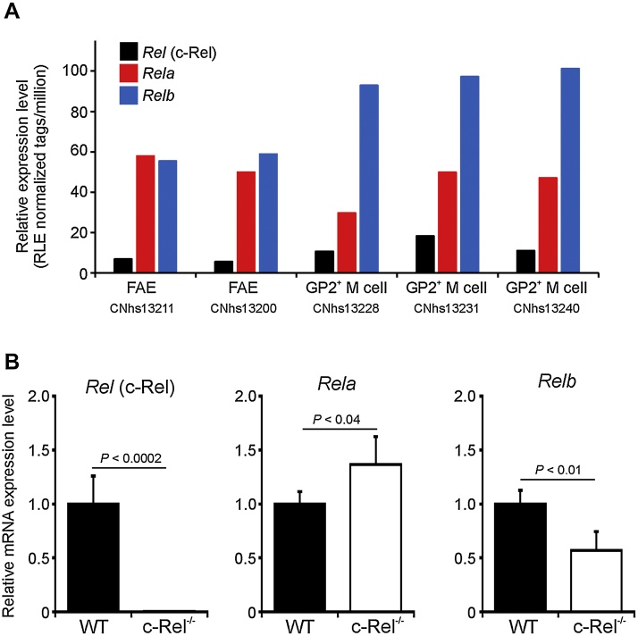
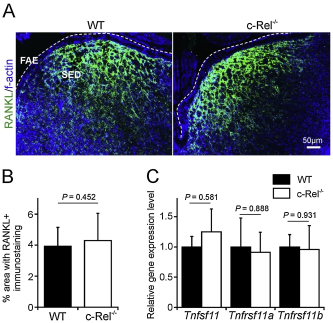
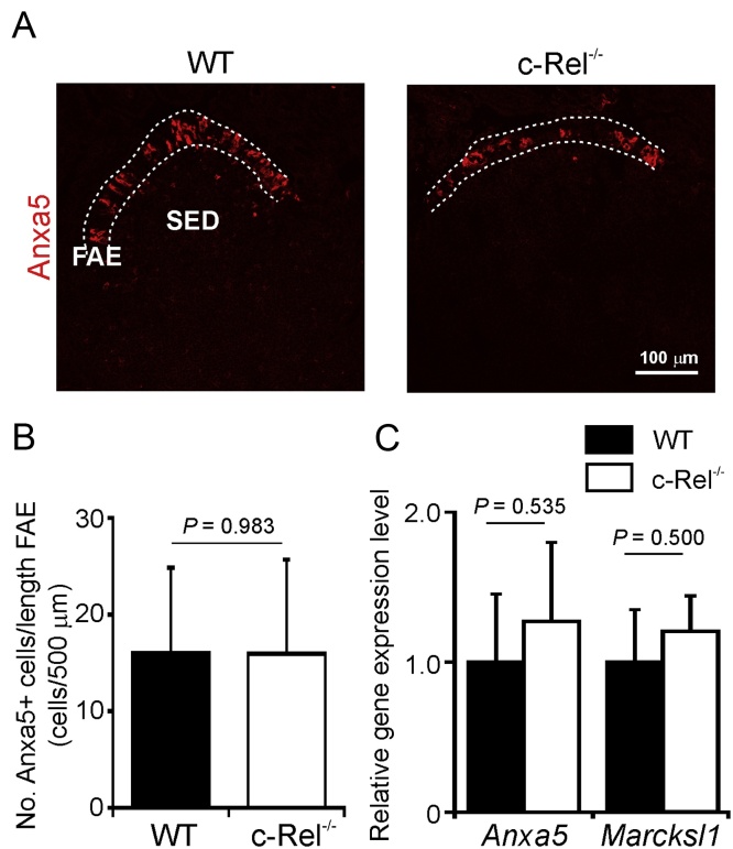
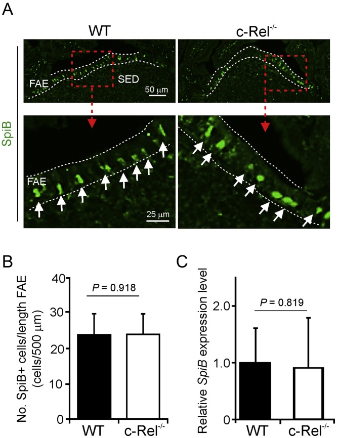
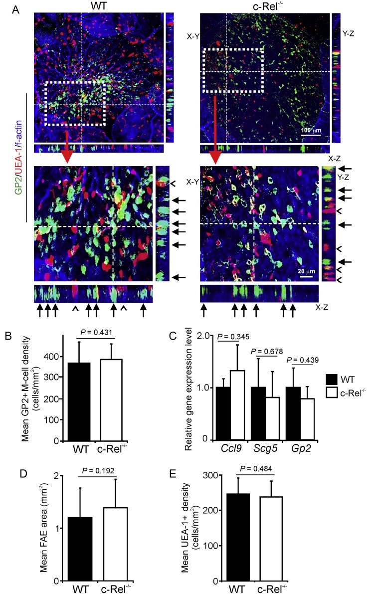
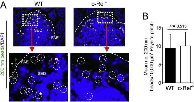
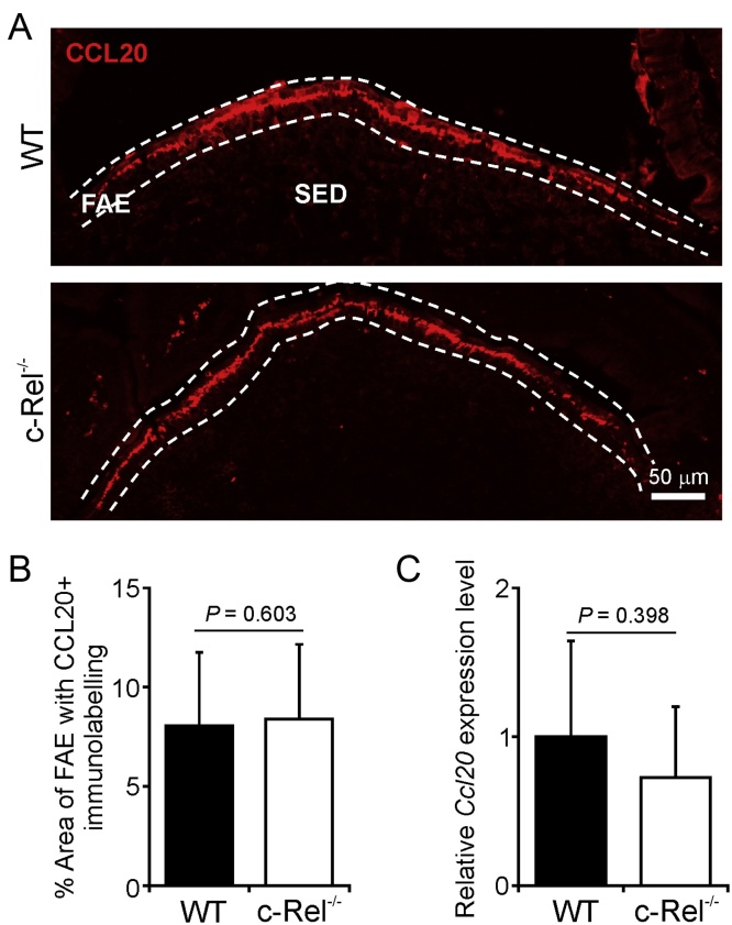
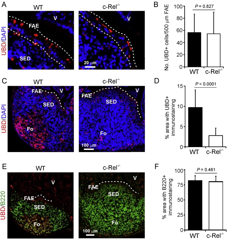
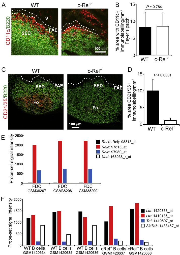
M cells reside within the follicle-associated epithelium (FAE) overlying the gut-associated lymphoid tissues. These unique phagocytic epithelial cells enable the mucosal immune system to sample antigens within the lumen of the intestine. The differentiation of M cells from uncommitted precursors in the FAE is dependent on the production of receptor activator of nuclear factor-κB ligand (RANKL) by subepithelial stromal cells. The ligation of a variety of cell surface receptors activates the nuclear factor-κB (NF-κB) family of transcription factors which in-turn induce the transcription of multiple target genes. RANKL-stimulation can stimulate the nuclear translocation of the NF-κB subunit c-Rel. We therefore used c-Rel-deficient mice to determine whether the differentiation and functional maturation of M cells in the Peyer's patches was dependent on c-Rel. Our data show that c-Rel-deficiency does not influence the expression of RANKL or RANK in Peyer's patches, or the induction of M-cell differentiation in the FAE. RANKL-stimulation in the differentiating M cells induces the expression of SpiB which is essential for their subsequent maturation. However, SpiB expression in the FAE was also unaffected in the absence of c-Rel. As a consequence, the functional maturation of M cells was not impaired in the Peyer's patches of c-Rel-deficient mice. Although our data showed that the specific expression of CCL20 and ubiquitin D in the FAE was not impeded in the absence of c-Rel, the expression of ubiquitin D was dramatically reduced in the B cell-follicles of c-Rel-deficient mice. Coincident with this, we also observed that the status of follicular dendritic cells in the B cell-follicles was dramatically reduced in Peyer's patches from c-Rel-deficient mice. Taken together, our data show that c-Rel is dispensable for the RANKL-mediated differentiation and functional maturation of M cells.

Keywords: Follicle-associated epithelium; M cells; Peyer’s patches; RANKL; c-Rel.


Figures

Fig. 1
Fig. 1
The NF-κB subunits c-Rel, RelA and RelB are expressed in Peyer’s patches in the FAE and M cells. (A) Comparison of Rel (which encodes c-Rel), Rela and Relb mRNA expression in deep CAGE RNA sequence data derived from individual samples of FAE and M cells in the FANTOM Consortium (Forrest et al., 2014) (http://fantom.gsc.riken.jp). (B) RT-qPCR analysis Rel, Rela and Relb expression in Peyer’s patches from c-Rel−/− or wild type (WT) control mice. Gene expression data are normalised so that the mean level in samples from WT mice was 1.0.
Fig. 2
Fig. 2
c-Rel-deficiency does not influence RANKL and RANK expression in Peyer’s patches. (A) IHC analysis suggested there was no observable difference in the expression or distribution of RANKL on sub-epithelial dome (SED) stromal cells in Peyer’s patches from c-Rel−/− and wild type (WT) control mice. Broken line indicates the lumenal surface of the follicle-associated epithelium (FAE). Sections are counterstained to detect f-actin (blue). (B) Morphometric analysis confirmed that the magnitude of RANKL-specific immunostaining observed in the SED of Peyer’s patches from WT and c-Rel−/− mice was similar. (C) RT-qPCR analysis suggested there was no significant difference in the expression levels of Tnfsf11 (RANKL), Tnfrsf11a (RANK) or Tnfrsf11b (osteoprotegerin) mRNA in Peyer’s patches from c-Rel−/− or WT mice. Gene expression data are normalised so that the mean level in samples from WT mice was 1.0. (For interpretation of the references to colour in this figure legend, the reader is referred to the web version of this article.)
Fig. 3
Fig. 3
c-Rel-deficiency does not influence expression of the immature M-cell markers Anxa5 and Marcksl1. (A) IHC analysis suggested there was no observable difference in the expression of Anxa5 in the FAE of Peyer’s patches from c-Rel−/− and wild type (WT) control mice. Broken lines indicate the boundaries of the FAE. (B) Morphometric analysis confirmed that the magnitude of Anxa5-specific immunostaining observed in the FAE of Peyer’s patches from c-Rel−/− and WT mice was similar. (C) RT-qPCR analysis suggested there was no significant difference in the expression levels of Anxa5 or Marcksl1 mRNA in Peyer’s patches from each mouse group. Gene expression data are normalised so that the mean level in WT was 1.0.
Fig. 4
Fig. 4
SpiB expression in the FAE is unaffected in the absence of c-Rel. (A) IHC analysis of SpiB expression (green) in the FAE of c-Rel−/− and wild-type (WT) control mice. Boxed areas in upper panels are shown at higher magnification in the lower panels. Broken lines indicate the FAE boundaries. Arrows, Spi-B+ cell nuclei in the FAE. (B) Morphometric analysis showed that the number of SpiB+ cells in the FAE of c-Rel−/− and WT were similar. (C) RT-qPCR analysis suggested there was no significant difference in the expression of Spib mRNA levels in Peyer’s patches from c-Rel−/− or WT mice. Gene expression data are normalised so that the mean level in samples from WT mice was 1.0. (For interpretation of the references to colour in this figure legend, the reader is referred to the web version of this article.)
Fig. 5
Fig. 5
c-Rel-deficiency does not influence the density of mature GP2+ M cells in the FAE. (A) Tissues were immunostained to detect GP2 (green), UEA-1 (red) and f-actin (blue). The positions of the X-Z and Y-Z projections of the FAE are indicated by the broken line in the main X-Y images. Closed arrows indicate GP2+ M cells with characteristic basolateral pockets. Open arrow-heads indicate GP2UEA-1+ goblet cells. The boxed areas in each of the main X-Y images are shown below at higher magnification. (B) Morphometric analysis revealed that the number of GP2+ M cells in the FAE of c-Rel−/− and wild type (WT) mice were similar (P = 0.431, two-tailed Mann-Whitney U test). (C) Comparison of the expression levels of Gp2, Ccl9 and Sgne1 mRNA in Peyer’s patches of c-Rel−/− and WT mice by RT-qPCR analysis. Gene expression data are normalised so that the mean level in WT mice was 1.0. (D) Morphometric analysis suggested that the size of the FAE in Peyer’s patches of c-Rel−/− and WT mice was similar (P = 0.192, two-tailed Mann-Whitney U test). (E) c-Rel-deficiency did not influence the density of GP2UEA-1+ goblet cells in the FAE (P = 0.484, two-tailed Mann-Whitney U test). (For interpretation of the references to colour in this figure legend, the reader is referred to the web version of this article.)
Fig. 6
Fig. 6
c-Rel deficiency does not influence the uptake of particulate antigen into the Peyer’s patches. (A) c-Rel−/− and wild-type (WT) mice were orally gavaged with 200 nm fluorescent microbeads and 24 h later, the presence of the microbeads in their Peyer’s patches was determined by fluorescence microscopical analysis. FAE, follicle-associated epithelium; SED, sub-epithelial dome; individual beads within the SED are highlighted by broken circles. (B) The number of beads transcytosed across the FAE of c-Rel−/− and WT mice was similar (P = 0.513, two-tailed Mann-Whitney U test). Data were collected from at least 10 sections from 3 to 5 Peyer’s patches from 4 to 7 mice/group.
Fig. 7
Fig. 7
c-Rel-deficiency does not influence CCL20 expression in the FAE. (a) IHC detection of CCL20 (red) in the FAE of c-Rel−/− and wild type (WT) control mice. Broken line indicates the boundaries of the FAE; SED, sub-epithelial dome. (b) Morphometric analysis showed that the magnitude of CCL20-specific immunostaining was similar in the FAE of c-Rel−/− and WT mice. (c) RT-qPCR analysis of Ccl20 mRNA levels in Peyer’s patches from c-Rel−/− or WT mice. Gene expression data are normalised so that the mean level in WT mice was 1.0. (For interpretation of the references to colour in this figure legend, the reader is referred to the web version of this article.)
Fig. 8
Fig. 8
Effect of c-Rel-deficiency on ubiquitin D (UBD) expression in Peyer’s patches. IHC detection of UBD (red) in (A) the follicle-associated epithelium (FAE) and (C) B cell follicles of Peyer’s patches from c-Rel−/− and wild type (WT) control mice. (B and D) Morphometric analysis of the magnitude of UBD-specific immunostaining in (B) the FAE, and (D) B cell follicles of Peyer’s patches from c-Rel−/− and WT mice. (E) IHC analysis of the distribution of B cells (B220+ cells, green) and UBD (red) in the Peyer’s patches of c-Rel−/− and WT mice. (F) Morphometric analysis of the magnitude of B220-specific immunostaining in Peyer’s patches from c-Rel−/− and WT mice. Broken lines indicates the boundary of the FAE. Fo, B-cell follicle; SED, subepithelial dome; V, villi. (For interpretation of the references to colour in this figure legend, the reader is referred to the web version of this article.)
Fig. 9
Fig. 9
Effect of c-Rel-deficiency on follicular dendritic cell (FDC) status in Peyer’s patches. (A) IHC of the distribution of CD11c+ cells (red) in the B cell-follicles (B220+ cells, green) of Peyer’s patches from c-Rel−/− and wild type (WT) control mice. (B) Morphometric analysis showed that the magnitude of CD11c-specific immunostaining was similar in tissues from c-Rel−/− and WT mice. (C) IHC analysis of FDC (CD21/35+ cells, red) in the B cell follicles (B220+ cells, green) of Peyer’s patches from c-Rel−/− and WT mice. (D) Morphometric analysis suggested that the magnitude of C21/35-specific immunostaining was dramatically reduced in tissues from c-Rel−/− mice when compared to WT mice. Broken lines in “A” and “C” indicate the boundary of the follicle-associated epithelium (FAE). Fo, B-cell follicle; SED, subepithelial dome; V, villi. (E) Expression of Rel, Rela, Relb and Ubd in publicly-available gene expression data of enriched-FDC performed on Affymetrix mouse genome U74v2 expression arrays (Huber et al., 2005). (F) Comparison of the expression of Lta, Ltb, Tnf and Slc7a6 in publicly-available gene expression data of c-Rel-deficient and WT B cells performed on Affymetrix mouse genome 430 2.0 expression arrays (Heise et al., 2014). The individual Affymetrix probe sets for each gene analysed are indicated. GSMXXXX, accession codes of each data set used. (For interpretation of the references to colour in this figure legend, the reader is referred to the web version of this article.)

Similar articles

Cited by

References

    1. Alcamo E., Hacohen N., Schulte L.C., Rennert P.D., Hynes R.O. Requirement for the NF-kappaB family member RelA in the development of secondary lymphoid organs. J. Exp. Med. 2002;195:233–244. - PMC - PubMed
    1. Bannard O., McGowan S.J., Ersching J., Ishido S., Victora G.D. Ubiquitin-mediated fluctations in MHC class II facilitate efficient germinal center B cell responses. J. Exp. Med. 2016;213:993–1009. - PMC - PubMed
    1. Barone F., Vossenkamper A., Boursier L., Su W., Watson A. IgA-producing plasma cells originate from germinal centres that are induced by B-cell receptor engagement in humans. Gastroenterology. 2011;140:947–956. - PMC - PubMed
    1. Bastide P., Darido C., Pannequin J., Kist R., Robine S. Sox9 regulates cell proliferation and is required for Paneth cell differentiation in the intestinal epithelium. J. Cell Biol. 2007;178:635–648. - PMC - PubMed
    1. Cho K.-J., Walseng E., Ishido S., Roche P.A. Ubiquitination by March-1 prevents MHC class II recycling and promotes MHC class II turnover in antigen-presenting cells. Proc. Natl. Acad. Sci. U. S. A. 2015;112:10449–10454. - PMC - PubMed

Publication types

MeSH terms