. 2016 Sep 23;13(1):251.
doi: 10.1186/s12974-016-0723-3.

Andrographolide induces Nrf2 and heme oxygenase 1 in astrocytes by activating p38 MAPK and ERK

Affiliations

Andrographolide induces Nrf2 and heme oxygenase 1 in astrocytes by activating p38 MAPK and ERK

Siew Ying Wong et al. J Neuroinflammation. .

Abstract

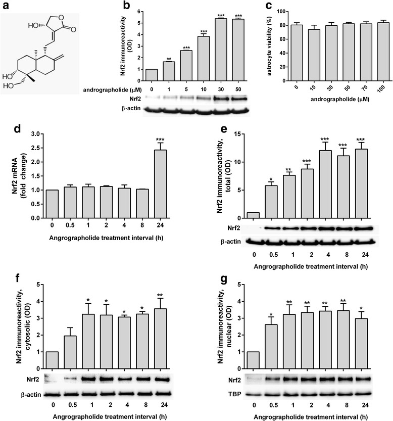
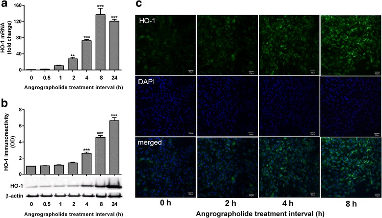
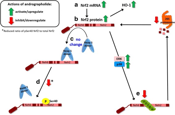
Background: Andrographolide is the major labdane diterpenoid originally isolated from Andrographis paniculata and has been shown to have anti-inflammatory and antioxidative effects. However, there is a dearth of studies on the potential therapeutic utility of andrographolide in neuroinflammatory conditions. Here, we aimed to investigate the mechanisms underlying andrographolide's effect on the expression of anti-inflammatory and antioxidant heme oxygenase-1 (HO-1) in primary astrocytes.

Methods: Measurements of the effects of andrograholide on antioxidant HO-1 and its transcription factor, Nrf2, include gene expression, protein turnover, and activation of putative signaling regulators.

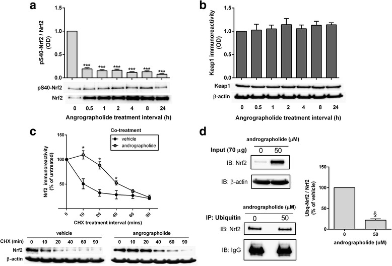
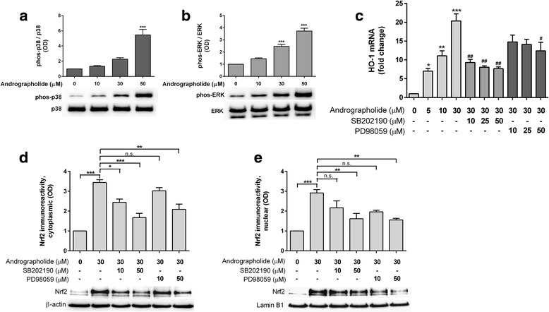
Results: Andrographolide potently activated Nrf2 and also upregulated HO-1 expression in primary astrocytes. Andrographolide's effects on Nrf2 seemed to be biphasic, with acute (within 1 h) reductions in Nrf2 ubiquitination efficiency and turnover rate, followed by upregulation of Nrf2 mRNA between 8 and 24 h. The acute regulation of Nrf2 by andrographolide seemed to be independent of Keap1 and partly mediated by p38 MAPK and ERK signaling.

Conclusions: These data provide further insights into the mechanisms underlying andrographolide's effects on astrocyte-mediated antioxidant, and anti-inflammatory responses and support the further assessment of andrographolide as a potential therapeutic for neurological conditions in which oxidative stress and neuroinflammation are implicated.

Keywords: Andrographolide; Antioxidant response; Astrocyte; Heme oxygenase 1; Mitogen-activated protein kinases; Nrf2.


Figures

Fig. 1
Fig. 1
Dose and time-course of andrographolide’s effects on Nrf2. a Structure of andrographolide: 3-[2-[decahydro-6-hydroxy-5-(hydroxymethyl)-5,8a-dimethyl-2-methylene-1-napthalenyl]ethylidene]dihydro-4-hydroxy-2(3H)-furanone (CAS no. 5508-58-7). b Bar graphs depict mean ± S.E.M. Nrf2 immunoreactivities (optical density, OD fold changes normalized to β-actin) in rat primary astrocytes treated with andrographolide at the indicated concentrations for 1 h (with representative immunoblot of 3 independent experiments), with the vehicle-only (“0 μM”) group set at 1. c Cell viability (in mean ± S.E.M. % of vehicle-only from three independent experiments) of primary astrocytes after 24 h treatment with andrographolide at the indicated concentrations. d mRNA and e immunoreactivity changes of Nrf2 in primary astrocytes treated with andrographolide (50 μM) for the indicated time intervals, together with immunoreactivites in the f cytosolic and g nuclear fractions (with representative immunoblots). Bar graphs show mRNA or immunoreactivity expressed as mean ± S.E.M. fold change in transcript level or optical density (OD), respectively, with vehicle-only (“0 h”) group set as 1, from three to four independent experiments. Raw transcript values were normalized to mean expression of housekeeping genes (see the “Methods” section) prior to conversion to fold-change values while immunoreactivities were normalized to β-actin except for Nrf2 nuclear fractions, which were normalized to TATA-binding protein (TBP). *p < 0.05; **p < 0.01; and ***p < 0.001; significantly different from vehicle-only group (one-way ANOVA with Dunnett’s post hoc tests)
Fig. 2
Fig. 2
Andrographolide increases stability of Nrf2 protein by altering ubiquitination efficiency. Primary astrocytes were treated with andrographolide (50 μM) for the indicated time intervals and measured for immunoreactivities of a pSer40 Nrf2/total Nrf2 as well as b Keap1 immunoreactivity normalized to β-actin (with representative immunoblots), and bar graphs showing mean ± S.E.M. fold changes in optical density (OD) with vehicle-only (“0 h”) group set as 1. ***p < 0.001; significantly different from vehicle-only group (one-way ANOVA with Dunnett’s post hoc tests). c Rat primary astrocytes were treated with cycloheximide (CHX, 10 μg/mL) with or without andrographolide (50 μM) co-incubation for the indicated time intervals and then measured for total Nrf2 immunoreactivity (with representative immunoblots). The graph represents mean ± S.E.M. Nrf2 immunoreactivities in vehicle-only (filled circles) and andrographolide co-treated (open circles) groups expressed as % of untreated (“0 h” CHX) group. *p < 0.05; significantly different from vehicle-only group (Student’s t tests). d Rat primary astrocytes were incubated with or without 50 μM of andrographolide for 1 h and processed for immunoprecipitation (see the “Methods” section), with representative input (IB) and ubiquitin-immunoprecipated (IP) blots for Nrf2. Bar graph shows mean ± S.E.M. immunoreactivity (fold changes in optical densities, OD with untreated group set at 100 %) of Ub-Nrf2/total Nrf2 normalized to β-actin. § p < 0.001 for comparison with untreated control (Student’s t tests). All data were from three to four independent experiments
Fig. 3
Fig. 3
Andrographolide upregulates heme oxygenase-1 in astrocytes. Primary astrocytes were treated with andrographolide (50 μM) for the indicated time intervals and measured for HO-1 a mRNA and b immunoreactivity (with representative immunoblots), together with respective bar graphs of mean ± S.E.M. fold changes in transcript level or optical density (OD), with vehicle-only (“0 h”) group set as 1, from three to four independent experiments. Raw transcript values were normalized to mean expression of housekeeping genes (see the “Methods” section) prior to conversion to fold-change values while HO-1 immunoreactivity was normalized to β-actin. **p < 0.01 and ***p < 0.001; significantly different from vehicle-only group (one-way ANOVA with Dunnett’s post hoc tests). c Primary astrocytes treated with 50 μM andrographolide for the indicated time intervals were processed for HO-1 immunofluorescence staining using Alexa Flour® 488-conjugated secondary antibody (green), while DAPI counter stain (blue) was used to visualize cell nuclei. Scale bars = 50 μm
Fig. 4
Fig. 4
Andrographolide induces HO-1 expression through p38 MAPK- and ERK-dependent regulation of Nrf2. Effects of andrographolide on a p38 MAPK and b ERK activation. Primary astrocytes were treated with increasing concentrations of andrographolide for 6 h and processed for immunobloting. Bar graphs showing mean ± S.E.M. fold changes in optical density (OD, with vehicle-only “0 μM” group set as 1) of phospho-protein normalized to total protein, ***p < 0.001; significantly different from vehicle-only group (one-way ANOVA with Dunnett’s post hoc tests). c Effects of p38 MAPK and ERK inhibition on HO-1 mRNA expression. Primary astrocytes were pretreated with or without SB202190 (p38 MAPK inhibitor) or PD98059 (MEK/ERK inhibitor) for an hour followed by 4 h of incubation with andrographolide (with presence of inhibitors) before processing for RT-PCR. Bar graphs are mean ± S.E.M. fold change in transcript level, with untreated group set as 1. Raw transcript values were normalized to mean expression of housekeeping genes (see the “Methods” section) prior to conversion to fold-change values. *p < 0.05, **p < 0.01, and ***p < 0.001; significantly difference from untreated group (one-way ANOVA followed by Dunnett’s post hoc tests.). # p < 0.05, ## p < 0.01; significantly different from 30 μM andrographolide (one- way ANOVA followed by Bonferroni’s post hoc tests). d, e Effects of p38 MAPK and ERK inhibition on Nrf2 in the cytosolic and nuclear compartments, respectively. Primary astrocytes were pretreated with PD98059 or SB202190 for 1 h followed by another 1 h of incubation with 30 μM andrographolide (in the presence of inhibitors). Cytoplasmic and nuclear fractions were separated and processed for immunoblotting. Bar graphs show immunoreactivities (mean ± S.E.M. fold changes in optical densities, OD, with untreated group set at 1) of Nrf2 normalized to β-actin (cytoplasmic) or to lamin B1 (nuclear). *p < 0.05, **p < 0.01, ***p < 0.001; significant pairwise difference and n.s. = not significant (p > 0.05) using one-way ANOVA with Bonferroni’s post hoc tests. All data were from three to four independent experiments
Fig. 5
Fig. 5
Andrographolide’s effects on Nrf2 and HO-1 in astrocytes. Summary schematic of findings of the current study. Black solid arrows indicate positive regulation, while activation effects of andrographolide are denoted by green arrows, and inhibitory effects are denoted by red arrows. a Andrographolide activates Nrf2 transcription between 8 and 24 h, leading to b increased Nrf2 proteins, which may also accumulate rapidly (within 1 h) through a process of altered ubiquitination which c does not alter Keap1 levels and d does not disrupt Nrf2-Keap1 binding through Ser40 phosphorylation within the Nrf2 Neh2 domain. Instead, andrographolide treatment leads to e reduced Nrf2 ubiquitination efficiency and subsequent 26S proteasomal turnover via p38 MAPK- and ERK-dependent pathways, although it is unclear whether the kinases directly act on Nrf2, or through regulation of other signaling molecules, for, e.g., GSK-3β. Further studies are required

Similar articles

Cited by

References

    1. Dandekar A, Mendez R, Zhang K. Cross talk between ER stress, oxidative stress, and inflammation in health and disease. Methods Mol Biol. 2015;1292:205–214. doi: 10.1007/978-1-4939-2522-3_15. - DOI - PubMed
    1. Freeman LC, Ting JP. The pathogenic role of the inflammasome in neurodegenerative diseases. J Neurochem. 2016;136 Suppl 1:29-38. - PubMed
    1. Dias V, Junn E, Mouradian MM. The role of oxidative stress in Parkinson’s disease. J Parkinsons Dis. 2013;3:461–491. - PMC - PubMed
    1. Lozano D, Gonzales-Portillo GS, Acosta S, de la Pena I, Tajiri N, Kaneko Y, Borlongan CV. Neuroinflammatory responses to traumatic brain injury: etiology, clinical consequences, and therapeutic opportunities. Neuropsychiatr Dis Treat. 2015;11:97–106. - PMC - PubMed
    1. Taylor JM, Main BS, Crack PJ. Neuroinflammation and oxidative stress: co-conspirators in the pathology of Parkinson’s disease. Neurochem Int. 2013;62:803–819. doi: 10.1016/j.neuint.2012.12.016. - DOI - PubMed

LinkOut - more resources