. 2016 Sep 22:5:31427.
doi: 10.3402/jev.v5.31427. eCollection 2016.

Extracellular vesicles are present in mouse lymph and their level differs in atherosclerosis

Affiliations

Extracellular vesicles are present in mouse lymph and their level differs in atherosclerosis

Andreea Milasan et al. J Extracell Vesicles. .

Abstract

The lymphatic system works in close collaboration with the cardiovascular system to preserve fluid balance throughout the body and is essential for the trafficking of antigen-presenting cells and lymphocytes to lymphoid organs. Recent findings have associated lymphatic dysfunction with the pathogenesis of cardiovascular-related diseases such as atherosclerosis, inflammation and obesity. Whether lymphatic dysfunction is a cause or a consequence of these diseases, as well as how, is under intensive investigation. Extracellular vesicles (EVs) are submicron vesicles released by diverse cell types upon activation or apoptosis and are considered important biomarkers for several inflammatory diseases. Thus, it is critical to characterize the presence of EVs in various biological tissues and fluids to delineate their origins and, subsequently, their functions. In the past few years, new techniques allowing the quantitative and qualitative analysis of EVs have emerged, thus facilitating the onset of studies bridging these vesicles to the lymphatic system. Using several state-of-the-art approaches, this article reports the presence of diverse EVs inclusively derived from red blood cells and platelets in lymph of healthy animals. Our results suggest that lymph from atherosclerotic mice displays a higher concentration of EVs, bringing forward the concept that EVs contained in lymph could either be a biomarker for lymphatic dysfunction or, conversely, for inflammatory disease progression.

Keywords: atherosclerosis; biomarkers; inflammation; lymphatic vessels; platelets.


Conflict of interest statement

and funding The authors have no other relevant affiliations or financial involvement with any organization or entity with a financial interest in or financial conflict with the subject matter or materials discussed in the manuscript apart from those disclosed.

Figures

Fig. 1
Fig. 1
Assessment of lymph purity following collection and processing. (a) Lymph was collected from the thoracic duct (dotted circle) on EDTA (0.1 M); following sample collection, cell counting was performed with a Nexcelom Auto X4 cell counter before (total lymph) and after (supernatant) centrifugation. Cells were stained with acridine orange (AO), a nuclear staining (nucleic acid binding) fluorescent dye that stains all nucleated cells, in whole blood and total lymph. The number of AO cells larger than 5 µM in diameter (i.e. red blood cells) was assessed using brightfield and fluorescence imaging for AO. (b) Total lymph was purposely contaminated at different concentrations (10−1, 10−2, 10−3) with whole blood (WB) and our sample of interest (lymph) was then standardized to WB and compared to all concentrations and lymph supernatant (control). (c) Total number of AO+ cells was detected in total lymph and lymph supernatant using fluorescence imaging for AO. (n=4 per group; *p<0.05, ***p<0.001).
Fig. 2
Fig. 2
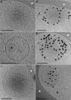
Representative cryo-electron microscopy images of lymph extracellular vesicles (EVs). (a through c) EVs devoid of annexin V gold nanoparticles indicate the absence of phosphatidylserine (PS) on their outer membrane surface. (d through f) EVs are labelled by annexin V gold nanoparticles, with the exception of the small unlabelled EVs marked with a black arrow. (b, d) The black asterisks point to circular shapes devoid of a lipid bilayer, which could most likely be lipoproteins. (a, c through e) The white asterisks point to areas of the supporting perforated carbon net. EVs devoid of annexin V gold nanoparticles (g) and EVs labelled by annexin V gold nanoparticles in the lymph of atherosclerotic LDLR-/- mice. Scale bars: 100 nm.
Fig. 3
Fig. 3
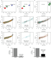
Detection of EVs in lymph using flow cytometry. (a through e) A Canto II flow cytometer modified with a “small particle” FSC PMT-H option was used to detect and quantify silica microspheres of 100 nm (red), 500 nm (green) and 1,000 nm (violet) mean diameter and EVs. (a, b) An EV gate was designed for the detection of small particles from 100 to 1,000 nm in diameter, based on the silica microspheres sizes (FSC PMT-H). (c) CD41+ mouse platelets are excluded from the EV gate based on their size (FSC PMT-H). Representative flow cytometry dot plot of lymph (d) total CFSE+ and (e) CFSE+PS+ EVs in control conditions or treated with 0.05% Triton or 50 M EDTA. Sensitivity to (f) Triton or (g) EDTA of EVs.
Fig. 4
Fig. 4
Dynamic light scattering size characterization of particles present in lymph of wild-type (WT) and LDLR-/- mice. Dimensions of the particles present in lymph from WT and LDLR-/- mice were determined with a Zetasizer Nano S, Malvern Instruments, UK Experiments were performed with 3 mice per experimental group.
Fig. 5
Fig. 5
Characterization of EVs in lymph of atherosclerotic mice. Flow cytometry was used to identify (a) total CFSE+ EVs based on their (b) PS expression. (c) CFSE+CD41+ and (e) CFSE+ Ter119+ EVs were analysed based on their (d, f) PS expression. Experiments were performed with 3 mice per experimental group. (*p<0.05, **p<0.01, ***p<0.001).

References

    1. Tammela T, Alitalo K. Lymphangiogenesis: molecular mechanisms and future promise. Cell. 2010;140:460–76. - PubMed
    1. Zolla V, Nizamutdinova IT, Scharf B, Clement CC, Maejima D, Akl T, et al. Aging-related anatomical and biochemical changes in lymphatic collectors impair lymph transport, fluid homeostasis, and pathogen clearance. Aging Cell. 2015;14:582–94. - PMC - PubMed
    1. Blum KS, Karaman S, Proulx ST, Ochsenbein AM, Luciani P, Leroux JC, et al. Chronic high-fat diet impairs collecting lymphatic vessel function in mice. PLoS One. 2014;9:e94713. - PMC - PubMed
    1. Lim HY, Rutkowski JM, Helft J, Reddy ST, Swartz MA, Randolph GJ, et al. Hypercholesterolemic mice exhibit lymphatic vessel dysfunction and degeneration. Am J Pathol. 2009;175:1328–37. - PMC - PubMed
    1. Vuorio T, Nurmi H, Moulton K, Kurkipuro J, Robciuc MR, Ohman M, et al. Lymphatic vessel insufficiency in hypercholesterolemic mice alters lipoprotein levels and promotes atherogenesis. Arterioscler Thromb Vasc Biol. 2014;34:1162–70. - PMC - PubMed