Review
. 2016 Oct;18(10):1338-49.
doi: 10.1093/neuonc/now182.

Tumor treating fields: a novel treatment modality and its use in brain tumors

Affiliations
Review

Tumor treating fields: a novel treatment modality and its use in brain tumors

Andreas F Hottinger et al. Neuro Oncol. 2016 Oct.

Abstract

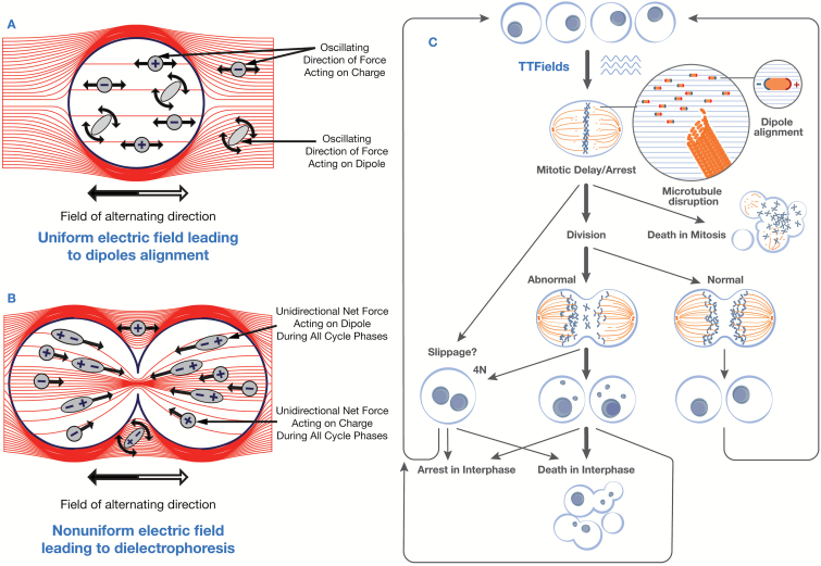
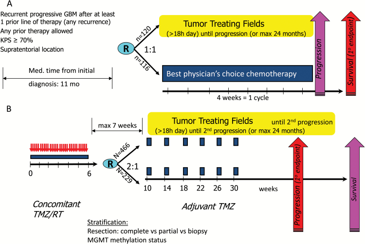
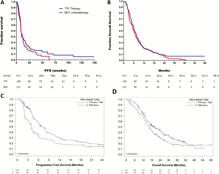
Tumor treating fields (TTFields) are low-intensity electric fields alternating at an intermediate frequency (200kHz), which have been demonstrated to block cell division and interfere with organelle assembly. This novel treatment modality has shown promise in a variety of tumor types. It has been evaluated in randomized phase 3 trials in glioblastoma (GBM) and demonstrated to prolong progression-free survival (PFS) and overall survival (OS) when administered together with standard maintenance temozolomide (TMZ) chemotherapy in patients with newly diagnosed GBM. TTFields are continuously delivered by 4 transducer arrays consisting each of 9 insulated electrodes that are placed on the patient's shaved scalp and connected to a portable device. Here we summarize the preclinical data and mechanism of action, the available clinical data, and further outlook of this treatment modality in brain tumors and other cancer indications.


Figures

Fig. 1.
Fig. 1.
Mechanisms of action of tumor treating fields in and around quiescent and dividing cells. Inside quiescent cells (1A), the field is uniform, and the oscillating electric forces result only in ‘‘vibration’’ of ions and dipoles (the forces associated with each half cycle are denoted with white and gray arrows). In contrast, the nonuniform field within dividing cells (1B) induces forces pushing all dipoles toward the furrow. [reprinted with permission from ref. 12] 1C: Tumor treating fields (TTFields) exert directional forces and result in abnormal spindle formation and subsequent mitotic arrest or delay, possibly due to improper attachment of chromosomes to the spindle fibers. Cells can die in mitotic arrest or progress to cell division leading to abnormal aneuploid progeny (highlighted by bold arrow). Abnormal daughter cells die in the subsequent interphase, undergo a permanent arrest or proliferate through additional mitosis where they will be subjected to further TTFields assault. [adapted with permission, refs. 11 and 12]
Fig. 2.
Fig. 2.
Tumor treating fields (TTFields) device (2nd generation Optune) and its clinical use TTFields are administered by 4 transducer arrays placed on the shaved scalp and connected to a portable device generating 200kHz electric fields within the brain. The position of the transducer arrays are determined by the localization of the tumor using a mapping software (NovoTalTM). (Photo with permission from the patient)
Fig. 3.
Fig. 3.
Design of the pivotal trials in GBM. A: EF-11 Trial Design B: EF-14 Trial Design
Fig. 4.
Fig. 4.
Progression-free and overall survival in EF-11 (A&B) & EF-14 (C&D) trials. EF-11 trial: Progression-free survival (A) and overall survival (B) of the intent-to-treat population. Hazard ratio for overall survival: 0.86 (CI, 0.66–1.12, P=0.66) [Reprinted with permission from ref, 16] Progression-free survival (C) and overall survival (D) of the intent-to-treat population in the EF-14 trial (interim data set). Hazard ratio for progression 0.63 (95%CI 0.43–0.89, P<0.01); for survival 0.74 (CI 0.56–0.98, P =0.0004). [Reprinted with permission, ref. 17]
Fig. 5.
Fig. 5.
Skin toxicities observed under tumor treating fields (TTFields). Some mild-moderate (grade 1–2) skin reaction is observed in up to half of patients (in EF-14 trial reported in 43%, grade 3 in 2%); however, it is usually self-limiting and resolves by removing the electrodes for a few days and applying local steroid-containing ointments. The images represent a few examples of skin reactions. (A) allergic contact dermatitis (B) irritant contact dermatitis (C) folliculitis (D), erosions [Reproduced from ref. 21].

References

    1. Palti Y. Stimulation of internal organs by means of externally applied electrodes. J Appl Physiol. 1966;21(5):1619–1623. - PubMed
    1. Storm FK, Morton DL, Kaiser LR, et al. Clinical radiofrequency hyperthermia: a review. Natl Cancer Inst Monogr. 1982;61:343–350. - PubMed
    1. Polk C, Postow E. Handbook of biological effects of electromagnetic fields. Boca Raton, FL: CRC Press; 1995.
    1. Takashima S, Schwan HP. Alignment of microscopic particles in electric fields and its biological implications. Biophys J. 1985;47(4):513–518. - PMC - PubMed
    1. Holzapfel C, Vienken J, Zimmermann U. Rotation of cells in an alternating electric field: theory and experimental proof. J Membr Biol. 1982;67(1):13–26. - PubMed