. 2017 Nov 1;27(11):5041-5053.
doi: 10.1093/cercor/bhw289.

N-Methyl d-Aspartate Receptor Expression Patterns in the Human Fetal Cerebral Cortex

Affiliations

N-Methyl d-Aspartate Receptor Expression Patterns in the Human Fetal Cerebral Cortex

Inseyah Bagasrawala et al. Cereb Cortex. .

Abstract

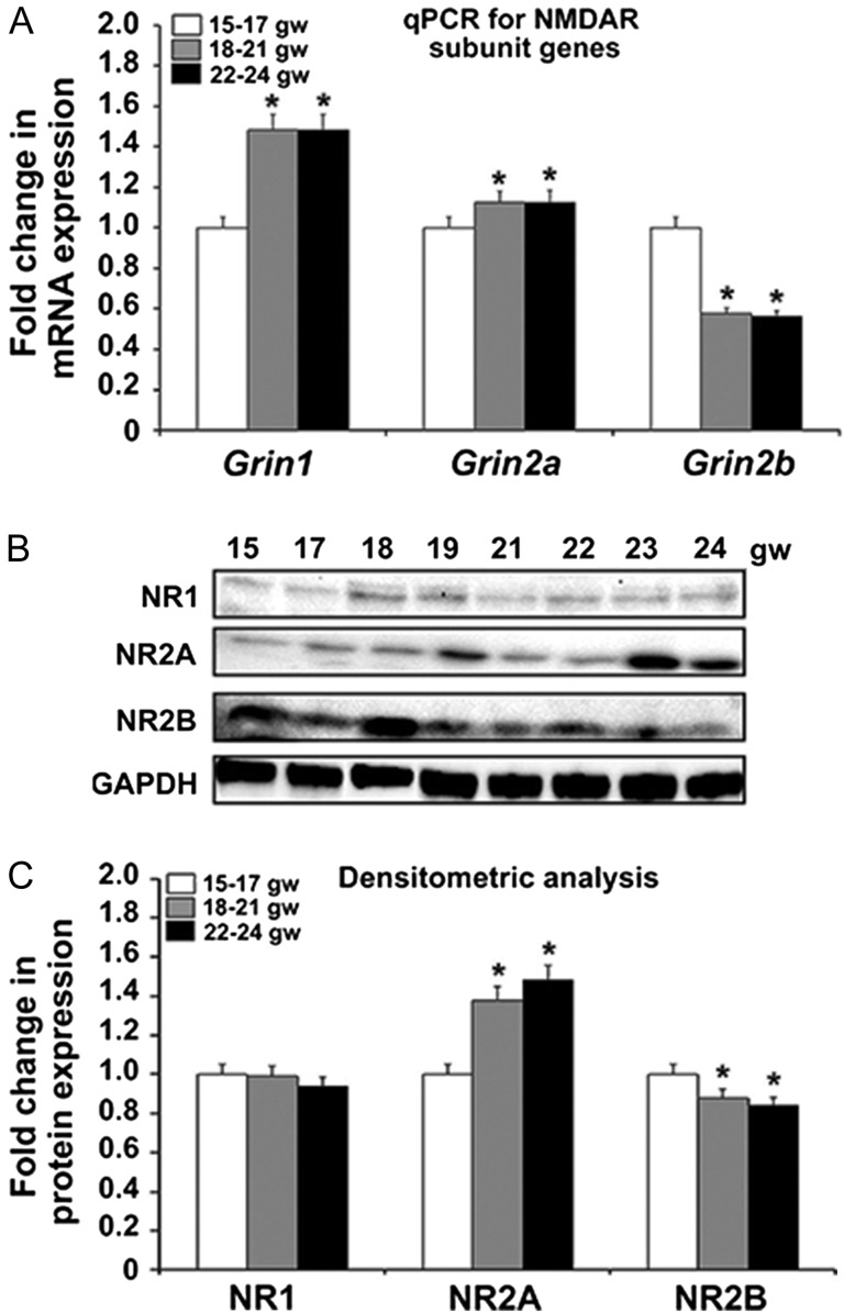
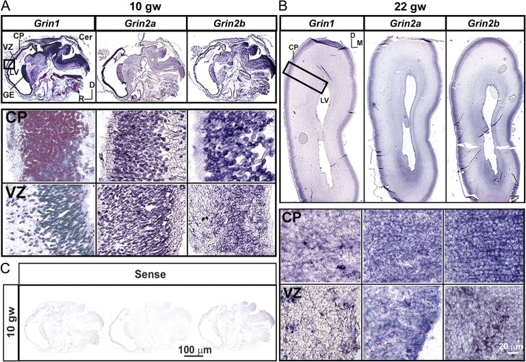
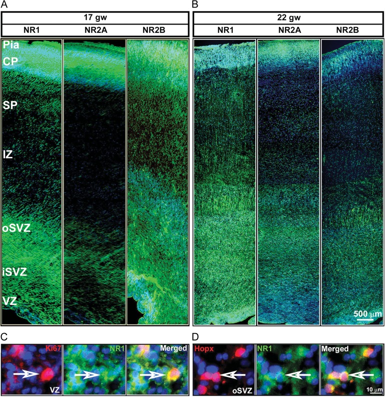
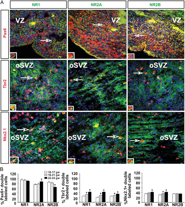
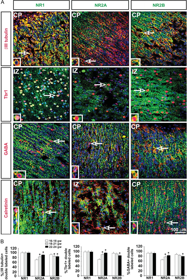
N-methyl d-aspartate receptors (NMDARs), a subtype of glutamate receptor, have important functional roles in cellular activity and neuronal development. They are well-studied in rodent and adult human brains, but limited information is available about their distribution in the human fetal cerebral cortex. Here we show that 3 NMDAR subunits, NR1, NR2A, and NR2B, are expressed in the human cerebral cortex during the second trimester of gestation, a period of intense neurogenesis and synaptogenesis. With increasing fetal age, expression of the NMDAR-encoding genes Grin1 (NR1) and Grin2a (NR2A) increased while Grin2b (NR2B) expression decreased. The protein levels of all 3 subunits paralleled the changes in gene expression. On cryosections, all 3 subunits were expressed in proliferative ventricular and subventricular zones, in radial glia, and in intermediate progenitor cells, consistent with their role in the proliferation of cortical progenitor cells and in the determination of their respective fates. The detection of NR1, NR2A, and NR2B in both glutamatergic and GABAergic neurons of the cortical plate suggests the involvement of NMDARs in the maturation of human cortical neurons and in early synapse formation. Our results and previous studies in rodents suggest that NMDAR expression in the developing human brain is evolutionarily conserved.

Keywords: glutamatergic receptors; human fetal development; immunohistochemistry; in situ hybridization.


Figures

Figure 1.
Figure 1.
The 3 NMDAR subunits, NR1, NR2A, and NR2B, are expressed in human fetal brain at midgestation. (A) Expression of the genes Grin1 and Grin2a increases with age, while that of Grin2b decreases from the youngest group 15–17 gw to the oldest group 22–24 gw (n = 3 per age group; P < 0.05). The mean values are plotted; the error bars represent ± the SEM. (B) Western blot of the NR1, NR2A, and NR2B subunits across all ages studied. GAPDH is the loading control. (C) Densitometric analysis confirms that NR1 protein expression does not change with age, while NR2A and NR2B protein levels parallel the trend in the expression of their respective genes, with an age-dependent increase in NR2A and decrease in NR2B (n = 3 per age group; P < 0.05). The mean values are plotted; the error bars represent ± the SEM.
Figure 2.
Figure 2.
Distributions of NR1, NR2A, and NR2B transcripts in the telencephalic wall. In situ hybridization reveals the presence of Grin1, Grin2a, and Grin2b mRNAs in the proliferative ventricular zone (VZ) and neuron-rich cortical plate (CP) as early as (A) 10 gw (sagittal section) and (B) at 22 gw (coronal section). Higher magnifications of the CP and VZ are shown in the insets. (C) Sense (control) probes for Grin1, Grin2a, and Grin2b shown for 10 gw. D-dorsal, M-medial, R-rostral. The whole sections images are captured by Aperio Versa (LeicaBiosystems.com), at ×4.
Figure 3.
Figure 3.
Immunohistochemistry of NR1, NR2A, and NR2B (green) along the various zones of the telencephalic wall of a representative case at 17 gw (A) and 22 gw (B). Greater immunolabeling intensity is seen for NR2A at 22 gw compared with 17gw, whereas the opposite is seen for NR2B subunit. (C) NR1 (green) expression on proliferative progenitors (Ki67+; red) in the VZ and (D) on basal radial glia (Hopx+; red) in the SVZ. Arrows point to double-labeled cells. Cell nuclei stained blue with bisbenzimide (BB).
Figure 4.
Figure 4.
Expression of NMDAR subunits on cortical progenitors. (A) Representative images of coronal cryosections at 17 gw. Double immunolabeling experiments with antibodies against the NR1, NR2A, and NR2B NMDAR subunits (green) and the progenitor subtypes (red) Pax6 (RGCs), Tbr2 (intermediate progenitors) and Nkx2.1 (interneuron progenitors) in the proliferative VZ/SVZ. Cell nuclei stained blue with BB. Boxed areas are presented on higher magnification in the inset. (B) Quantification of the percentage of Pax6+, Tbr2+, and Nkx2.1+ progenitors expressing the 3 NMDAR subunits in the 3 age groups (n = 3 per age group; P < 0.05); The mean values are plotted; the error bars represent ± the SEM.
Figure 5.
Figure 5.
Expression of NMDAR subunits on cortical neurons. (A) Representative images at 17 gw. Double immunolabeling experiments with antibodies against the NR1, NR2A, and NR2B NMDAR subunits (green) and the neuronal subtypes (red) βIII tubulin (neurons), Tbr1 (glutamatergic), GABA (interneurons), and calretinin (interneuron subtype) in the cortical plate (CP) and the transient intermediate zone (IZ). Cell nuclei stained blue with BB. Arrows point to cells presented on higher magnification in the inset. (B) Quantification of the percentage of βIII tubulin+ neurons, Tbr1+ glutamatergic neurons, and GABA+ interneurons expressing the 3 NMDAR subunits in the 3 age groups studied (n = 3 per age group; P < 0.05). The mean values are plotted; the error bars represent ± the SEM.

References

    1. Arnsten AF, Wang MJ, Paspalas CD. 2012. Neuromodulation of thought: flexibilities and vulnerabilities in prefrontal cortical network synapses. Neuron. 76:223–239. - PMC - PubMed
    1. Balasz R. 2006. Trophic effect of glutamate. Curr Top Med Chem. 6:961–968. - PubMed
    1. Barria A, Malinow R. 2002. Subunit-specific NMDA receptor trafficking to synapses. Neuron. 35:345–353. - PubMed
    1. Barria A, Malinow R. 2005. NMDA receptor subunit composition controls synaptic plasticity by regulating binding to CaMKII. 48:289–301. - PubMed
    1. Bayatti N, Moss JA, Sun L, Ambrose P, Ward JF, Lindsay S, Clowry GJ. 2008. A molecular neuroanatomical study of the developing human neocortex from 8 to 17 postconceptional weeks revealing the early differentiation of the subplate and subventricular zone. Cereb Cortex. 18:1536–1548. - PMC - PubMed

Publication types

MeSH terms