

The use of blocking screws with internal lengthening nail and reverse rule of thumb for blocking screws in limb lengthening and deformity correction surgery
- PMID: 27665618
- PMCID: PMC5069203
- DOI: 10.1007/s11751-016-0265-3
The use of blocking screws with internal lengthening nail and reverse rule of thumb for blocking screws in limb lengthening and deformity correction surgery
Abstract
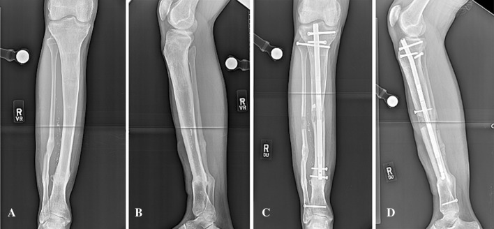
Internal lengthening nail (ILN) is a recent development in limb lengthening and deformity correction specialty. The ILN has the distinct advantage of combining acute deformity correction with gradual lengthening of bone. While using ILN, the short metaphyseal bone fragment may develop a deformity at the time of osteotomy and nail insertion or during bone lengthening because of the wide medullary canal. These deformities are typically predictable, and blocking screws (Poller screws) are helpful in these situations. This manuscript describes the common deformities that occur in femur and tibia with osteotomies at different locations while using ILN in antegrade and retrograde nailing technique. Also, a systematic approach to the appropriate use of blocking screws in these deformities is described. In addition, the "reverse rule of thumb" is introduced as a quick reference to determine the ideal location(s) and number of blocking screws. These principles are applicable to limb lengthening and deformity correction as well as fracture fixation using intramedullary nails.
Keywords: Blocking screws; Deformity correction; Intramedullary lengthening nail; Limb lengthening; Reverse rule of thumb.
Conflict of interest statement
Saravanaraja Muthusamy declares no conflict of interest. S Robert Rozbruch, is a consultant with and receives payment for lectures including service on speakers’ bureaus from Smith and Nephew and receives royalties from Small Bone Innovations. Austin T. Fragomen is a consultant with Synthes and Smith and Nephew and receives payment for lectures including service on speakers’ bureaus from Smith and Nephew. Statement of human and animal rights All procedures performed in studies involving human participants were in accordance with the ethical standards of the institutional and/or national research committee and with the 1964 Helsinki declaration and its later amendments or comparable ethical standards. Informed consent For this kind of study formal consent is not required.
Figures
Similar articles
-
Blocking Screw-assisted Intramedullary Nailing Using the Reverse-rule-of-thumbs for Limb Lengthening and Deformity Correction.Strategies Trauma Limb Reconstr. 2019 May-Aug;14(2):77-84. doi: 10.5005/jp-journals-10080-1430. Strategies Trauma Limb Reconstr. 2019. PMID: 32742418 Free PMC article.
-
Acute Correction of Multiplanar Proximal Tibial Deformity Utilizing Fixator-Assisted Intramedullary Nailing.JBJS Essent Surg Tech. 2022 Aug 31;12(3):e21.00045. doi: 10.2106/JBJS.ST.21.00045. eCollection 2022 Jul-Sep. JBJS Essent Surg Tech. 2022. PMID: 36816522 Free PMC article.
-
Blocking screws for alignment control in intramedullary limb lengthening.Injury. 2017 Jul;48(7):1597-1602. doi: 10.1016/j.injury.2017.03.043. Epub 2017 Mar 31. Injury. 2017. PMID: 28381356
-
Retrograde magnetic internal lengthening nail for acute femoral deformity correction and limb lengthening.Expert Rev Med Devices. 2017 Oct;14(10):811-820. doi: 10.1080/17434440.2017.1378092. Epub 2017 Sep 17. Expert Rev Med Devices. 2017. PMID: 28893094 Review.
-
Intramedullary lengthening nails: can we also correct deformities?J Child Orthop. 2016 Dec;10(6):511-516. doi: 10.1007/s11832-016-0782-0. Epub 2016 Nov 15. J Child Orthop. 2016. PMID: 27848194 Free PMC article. Review.
Cited by
-
A Comparison of Femoral Lengthening Methods Favors the Magnetic Internal Lengthening Nail When Compared with Lengthening Over a Nail.HSS J. 2018 Jul;14(2):166-176. doi: 10.1007/s11420-017-9596-y. Epub 2018 Jan 5. HSS J. 2018. PMID: 29983659 Free PMC article.
-
Regenerate Deformity with the Precice Tibial Nail.Strategies Trauma Limb Reconstr. 2020 May-Aug;15(2):98-105. doi: 10.5005/jp-journals-10080-1457. Strategies Trauma Limb Reconstr. 2020. PMID: 33505526 Free PMC article.
-
Simultaneous correction of leg length discrepancy and angular deformity of the distal femur with retrograde Precice nails: a retrospective analysis of 45 patients.Acta Orthop. 2024 Jul 15;95:364-372. doi: 10.2340/17453674.2024.40947. Acta Orthop. 2024. PMID: 39007719 Free PMC article.
-
Bone Ninja Mobile App for Reverse Planning Method in Internal Limb Deformity and Lengthening Surgery.Strategies Trauma Limb Reconstr. 2019 May-Aug;14(2):72-76. doi: 10.5005/jp-journals-10080-1425. Strategies Trauma Limb Reconstr. 2019. PMID: 32742417 Free PMC article.
-
Total hip arthroplasty and femoral nail lengthening for hip dysplasia and limb-length discrepancy.Arthroplast Today. 2018 May 3;4(3):279-286. doi: 10.1016/j.artd.2018.03.001. eCollection 2018 Sep. Arthroplast Today. 2018. PMID: 30186905 Free PMC article.
References
-
- Krettek C, Miclau T, Schandelmaier P, Stephan C, Möhlmann U, Tscherne H. The mechanical effect of blocking screws (“Poller screws”) in stabilizing tibia fractures with short proximal or distal fragments after insertion of small-diameter intramedullary nails. J Orthop Trauma. 1999;13:550–553. doi: 10.1097/00005131-199911000-00006. - DOI - PubMed
LinkOut - more resources
Full Text Sources
Other Literature Sources
