. 2016 Sep 26:6:34043.
doi: 10.1038/srep34043.

Paraspeckles modulate the intranuclear distribution of paraspeckle-associated Ctn RNA

Affiliations

Paraspeckles modulate the intranuclear distribution of paraspeckle-associated Ctn RNA

Aparna Anantharaman et al. Sci Rep. .

Abstract

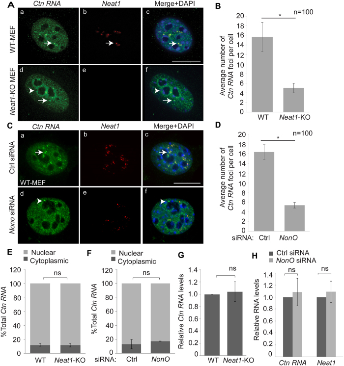
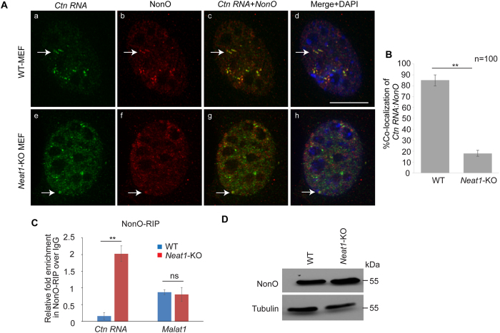
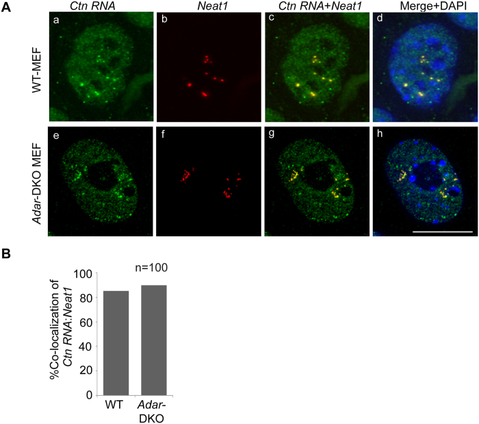
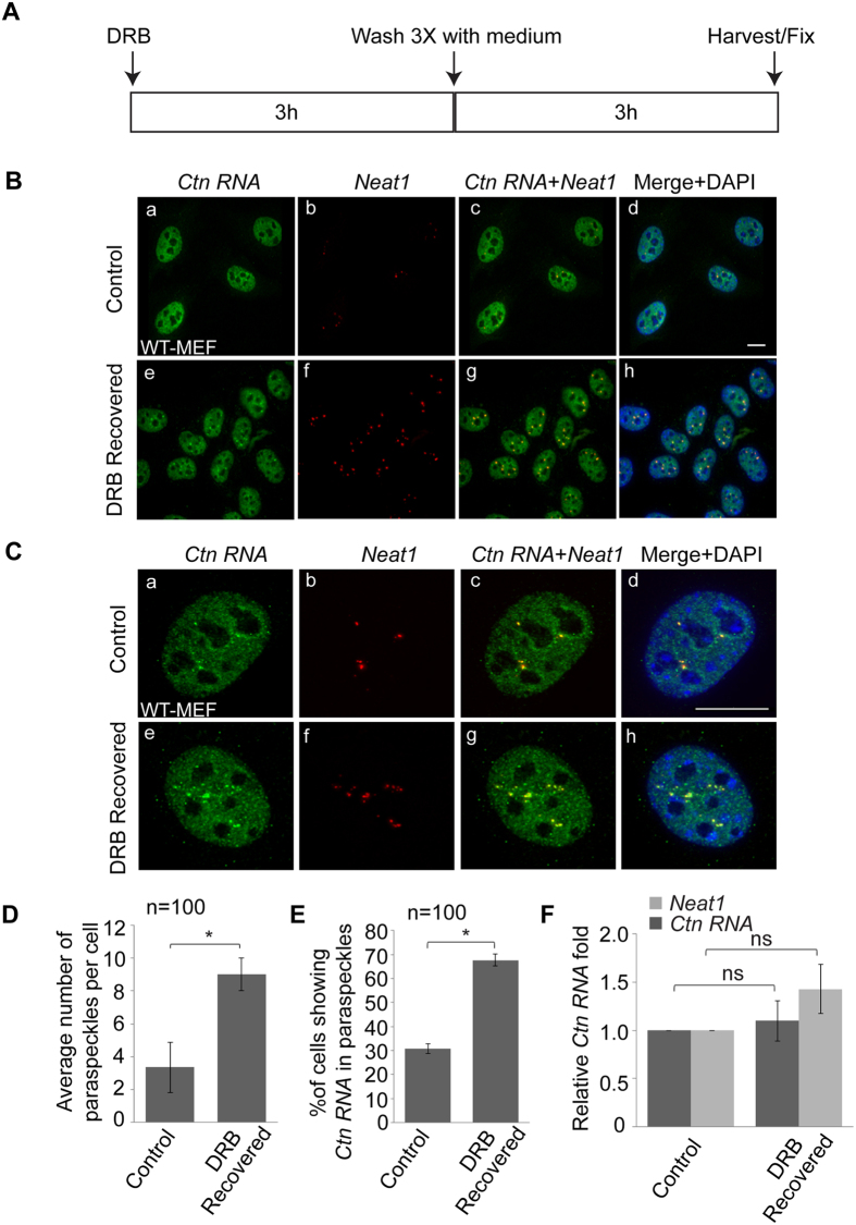
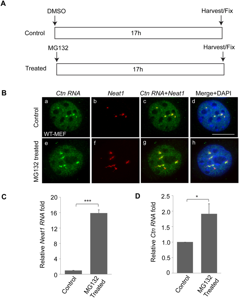
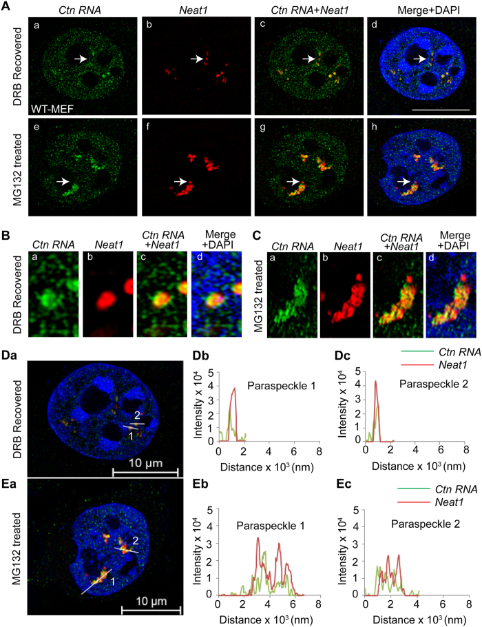
Paraspeckles are sub-nuclear domains that are nucleated by long noncoding RNA Neat1. While interaction of protein components of paraspeckles and Neat1 is understood, there is limited information on the interaction of non-structural RNA components with paraspeckles. Here, by varying paraspeckle number and size, we investigate how paraspeckles influence the nuclear organization of their non-structural RNA component Ctn RNA. Our results show that Ctn RNA remains nuclear-retained in the absence of intact paraspeckles, suggesting that they do not regulate nuclear retention of Ctn RNA. In the absence of Neat1, Ctn RNA continues to interact with paraspeckle protein NonO to form residual nuclear foci. In addition, in the absence of Neat1-nucleated paraspeckles, a subset of Ctn RNA localizes to the perinucleolar regions. Concomitant with increase in number of paraspeckles, transcriptional reactivation resulted in increased number of paraspeckle-localized Ctn RNA foci. Similar to Neat1, proteasome inhibition altered the localization of Ctn RNA, where it formed enlarged paraspeckle-like foci. Super-resolution structured illumination microscopic analyses revealed that in paraspeckles, Ctn RNA partially co-localized with Neat1, and displayed a more heterogeneous intra-paraspeckle localization. Collectively, these results show that while paraspeckles do not influence nuclear retention of Ctn RNA, they modulate its intranuclear compartmentalization.


Figures

Figure 1
Figure 1. Ctn RNA is nuclear-retained in absence of intact paraspeckles.
(A) RNA-FISH to detect Ctn RNA (green) and Neat1 (red) in DRB-recovered (3 hrs) WT and Neat1-KO MEFs. Arrow (a–f) indicates Ctn RNA foci and arrowhead (d,f) indicates perinucleolar localization of Ctn RNA. (B) Graph showing average number of Ctn RNA foci per cell in WT and Neat1-KO MEFs. (C) RNA-FISH to detect Ctn RNA and Neat1 in DRB-recovered control (Ctrl) and NonO-depleted transformed WT-MEFs. Please note that Ctn RNA shows increased paraspeckle association upon DRB recovery (please see Fig. 4). Arrow (a,c) marks Ctn RNA and Neat1 positive paraspeckle. Arrowhead (d,f) shows Ctn RNA positive but Neat1 negative paraspeckle-like nuclear body. (D) Graph showing average number of Ctn RNA foci per cell in (Ctrl) and NonO-depleted transformed WT-MEFs. (E,F) RT-qPCR to estimate Ctn RNA levels in nuclear and cytoplasmic fractions of (E) WT and Neat1-KO MEFs and (F) Ctrl and NonO-depleted MEFs. (G) RT-qPCR to detect total levels of Ctn RNA in WT and Neat1-KO MEFs. (H) Total levels of Ctn RNA in control and NonO siRNA treated MEFs. 3′UTR-1 primer pair has been used to measure Ctn RNA levels (Figure S2). Gapdh was used as the normalization control in RT-qPCR experiments. Scale bar indicates 10 μm. Error bars in (B,D,E–H) represent mean ± SD of three independent experiments. *P < 0.05, ns: not significant, using Student’s t test.
Figure 2
Figure 2. Ctn RNA interacts with paraspeckle component NonO in absence of intact paraspeckles.
(A) RNA-FISH to detect Ctn RNA (green) and NonO (red) in DRB-recovered WT and Neat1-KO MEFs. Scale bar indicates 10 μm. Arrow (a–h) indicates Ctn RNA and NonO positive nuclear foci. DNA is counterstained with DAPI (blue). (B) Graph showing percentage co-localization in WT and Neat1-KO MEFs. (C) NonO-RIP (RNA immunoprecipitation) followed by RT-qPCR analysis of Ctn RNA to determine interaction of NonO and Ctn RNA in WT and Neat1-KO MEFs. (D) Western blot showing NonO levels in WT and Neat1-KO MEFs. Tubulin was used as a loading control. Gapdh was used as the normalization control in RT-qPCR experiments. Error bars in (B,C) represent mean ± SD of three independent experiments. **P < 0.01 using Student’s t test.
Figure 3
Figure 3. ADARs do not influence the association of A-to-I edited Ctn RNA to paraspeckles.
(A) RNA-FISH to detect Ctn RNA (green) and Neat1 (red) in DRB-recovered WT and Adar1/Adar2 double knockout-KO (DKO) MEFs. DNA is counterstained with DAPI (blue). Scale bar indicates 10 μm. (B) % co-localization of Ctn RNA and Neat1 in the paraspeckles of DRB-recovered WT and Adar1/Adar2 double knockout-KO (DKO) MEFs.
Figure 4
Figure 4. Number of Ctn RNA positive foci and their association with paraspeckles is increased upon transcriptional reactivation.
(A) Schematic showing the experimental design. (B) RNA-FISH to detect Ctn RNA (green) and Neat1 (red) in control (ethanol) and DRB-recovered transformed WT-MEFs. (C) RNA-FISH analysis of Ctn RNA and Neat1 localization in a single cell of control (ethanol-treated) and DRB-recovered transformed WT-MEF. (D) Graph showing average number of paraspeckles per cell in control (ethanol-treated) and DRB-recovered transformed WT-MEFs. (E) Graph showing percentage of cells positive for Ctn RNA foci in control (ethanol-treated) and DRB-recovered transformed WT-MEFs. (F) RT-qPCR analysis of Ctn RNA levels in control (ethanol-treated) and DRB-recovered transformed WT-MEFs. Gapdh was used as the normalization control in RT-qPCR experiments. Scale bar indicates 10 μm. DNA is counterstained with DAPI (blue). Error bars in (D–F) represent mean ± SD of three independent experiments. *P < 0.05, ns: not significant using Student’s t test.
Figure 5
Figure 5. Ctn RNA forms enlarged foci in proteasome-inhibited cells.
(A) Schematic showing the experimental design. (B) RNA-FISH analysis of Ctn RNA (green) and Neat1 (red) localization in control (DMSO-treated) and MG132-treated transformed WT-MEFs. Scale bar indicates 10 μm. DNA is counterstained with DAPI (blue). (C) RT-qPCR analysis of Neat1 RNA levels in control (DMSO-treated) and MG132-treated transformed WT-MEFs. (D) RT-qPCR analysis of Ctn RNA levels in control (DMSO-treated) and MG132-treated transformed WT-MEFs. Gapdh was used as the normalization control in RT-qPCR experiments. Error bars in (C,D) represent mean ± SD of three independent experiments. ***P < 0.001, *P < 0.05 using Student’s t test.
Figure 6
Figure 6. Intra-paraspeckle localization of Ctn RNA.
(A) Super-resolution structured illumination microscopy (SR-SIM) of Ctn RNA (green) and Neat1 (red) localization in DRB recovered and MG132-treated transformed WT-MEFs. Scale bar indicates 10 μm. Arrow (a–h) indicates paraspeckle where Ctn RNA does not show co-localization with Neat1. (B,C) Co-localization of a single paraspeckle in (B) DRB recovered and (C) MG132-treated transformed WT-MEF. (D,E) Quantitation of Ctn RNA and Neat1 co-localization in (Da–c) DRB recovered and (Ea–c) MG132 treated transformed WT-MEFs (performed using ZEN 2012). Numbers in image indicate the specific paraspeckle analyzed and corresponds to the number mentioned in the graph. For example, “1” in image refers to “paraspeckle 1” in graph.

References

    1. Matera A. G., Izaguire-Sierra M., Praveen K. & Rajendra T. K. Nuclear bodies: random aggregates of sticky proteins or crucibles of macromolecular assembly? Dev Cell 17, 639–647, 10.1016/j.devcel.2009.10.017 (2009). - DOI - PMC - PubMed
    1. Caudron-Herger M. & Rippe K. Nuclear architecture by RNA. Curr Opin Genet Dev 22, 179–187, 10.1016/j.gde.2011.12.005 (2012). - DOI - PubMed
    1. Dundr M. & Misteli T. Biogenesis of nuclear bodies. Cold Spring Harb Perspect Biol 2, a000711, 10.1101/cshperspect.a000711 (2010). - DOI - PMC - PubMed
    1. Mao Y. S., Zhang B. & Spector D. L. Biogenesis and function of nuclear bodies. Trends Genet 27, 295–306, 10.1016/j.tig.2011.05.006 (2011). - DOI - PMC - PubMed
    1. Spector D. L. & Lamond A. I. Nuclear speckles. Cold Spring Harb Perspect Biol 3, 10.1101/cshperspect.a000646 (2011). - DOI - PMC - PubMed

LinkOut - more resources