

Platelet-rich plasma protects rat chondrocytes from interleukin-1β-induced apoptosis
- PMID: 27665780
- PMCID: PMC5101884
- DOI: 10.3892/mmr.2016.5767
Platelet-rich plasma protects rat chondrocytes from interleukin-1β-induced apoptosis
Abstract
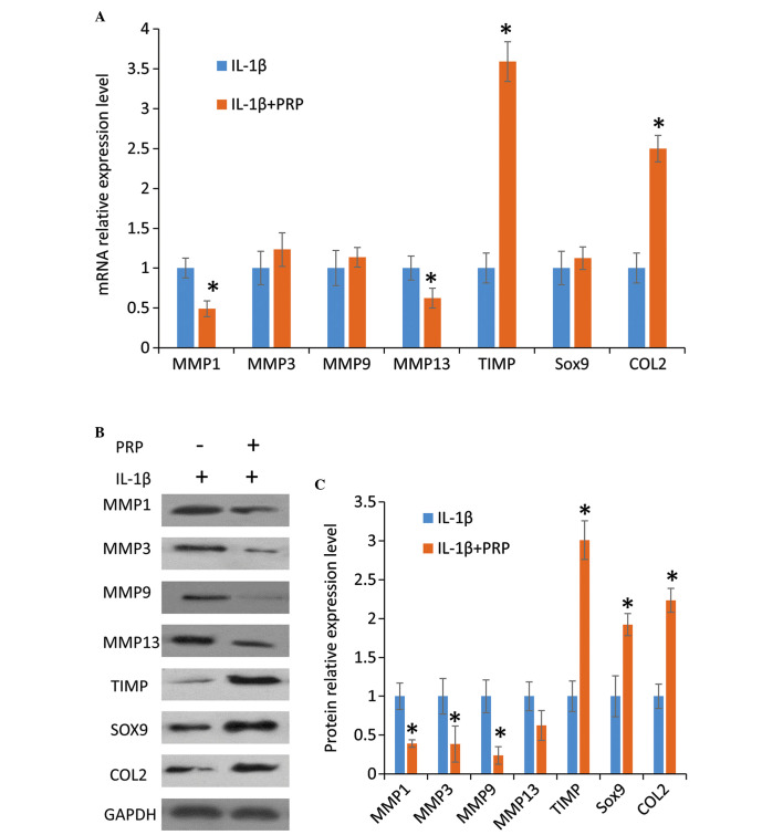
Interleukin (IL)-1β-induced chondrocyte apoptosis is associated with the pathogenesis of arthritis. Platelet‑rich plasma (PRP), which is derived from the patient's own blood and contains numerous growth factors, has the potential for arthritis treatment. Therefore, the present study aimed to determine the effects of PRP on chondrocyte apoptosis, under IL‑1β‑induced pathological conditions. Chondrocytes isolated from the knee joint of Sprague Dawley rats were used in the present study. Cell viability was determined using the Cell Counting kit‑8 assay, cell apoptosis was evaluated by flow cytometry, and the expression of apoptosis‑, anabolism‑ and catabolism-associated genes were detected by quantitative polymerase chain reaction; protein expression was detected by western blot analysis. The results demonstrated that 10% PRP in the culture medium increased chondrocyte proliferation, whereas IL‑1β induced cell apoptosis. Treatment with PRP significantly attenuated cell apoptosis in IL‑1β‑treated chondrocytes, and altered apoptosis‑associated expression at the gene and protein level. Furthermore, treatment with PRP significantly reduced matrix metalloproteinase production and promoted anabolism of cartilage extracellular matrix under IL‑1β treatment. The present study demonstrated the protective effects of PRP on chondrocyte apoptosis and extracellular matrix anabolism, and provided scientific evidence to support the potential use of PRP as a promising therapeutic strategy for the treatment of arthritis.
Figures


Similar articles
-
Platelet-rich plasma attenuates interleukin-1β-induced apoptosis and inflammation in chondrocytes through targeting hypoxia-inducible factor-2α.Tissue Cell. 2021 Dec;73:101646. doi: 10.1016/j.tice.2021.101646. Epub 2021 Sep 8. Tissue Cell. 2021. PMID: 34536814
-
Anti-Inflammatory Effects of Novel Standardized Platelet Rich Plasma Releasates on Knee Osteoarthritic Chondrocytes and Cartilage in vitro.Curr Pharm Biotechnol. 2019;20(11):920-933. doi: 10.2174/1389201020666190619111118. Curr Pharm Biotechnol. 2019. PMID: 31237204
-
Platelet-rich plasma inhibits Wnt/β-catenin signaling in rabbit cartilage cells activated by IL-1β.Int Immunopharmacol. 2018 Feb;55:282-289. doi: 10.1016/j.intimp.2017.12.031. Epub 2017 Dec 30. Int Immunopharmacol. 2018. PMID: 29291543
-
Platelet-rich plasma alleviates knee arthritis in rats by inhibiting p65.Cell Tissue Bank. 2024 Jun;25(2):463-473. doi: 10.1007/s10561-023-10102-3. Epub 2023 Jul 27. Cell Tissue Bank. 2024. PMID: 37501011
-
Platelet-rich plasma in the pathologic processes of cartilage: review of basic science evidence.Arthroscopy. 2013 Aug;29(8):1399-409. doi: 10.1016/j.arthro.2013.03.004. Epub 2013 May 11. Arthroscopy. 2013. PMID: 23669235 Review.
Cited by
-
Management of Hepple Stage V Osteochondral Lesion of the Talus with a Platelet-Rich Plasma Scaffold.Biomed Res Int. 2017;2017:6525373. doi: 10.1155/2017/6525373. Epub 2017 Mar 16. Biomed Res Int. 2017. PMID: 28401159 Free PMC article.
-
Platelet-rich Plasma-induced Inhibition of Chondrocyte Apoptosis Directly Affects Cartilage Thickness in Osteoarthritis.Cureus. 2019 Nov 1;11(11):e6050. doi: 10.7759/cureus.6050. Cureus. 2019. PMID: 31827985 Free PMC article.
-
The role of WNT and IL-1 signaling in osteoarthritis: therapeutic implications for platelet-rich plasma therapy.Front Aging. 2023 Jun 8;4:1201019. doi: 10.3389/fragi.2023.1201019. eCollection 2023. Front Aging. 2023. PMID: 37362206 Free PMC article. Review.
-
Cell and Cell Free Therapies in Osteoarthritis.Biomedicines. 2021 Nov 19;9(11):1726. doi: 10.3390/biomedicines9111726. Biomedicines. 2021. PMID: 34829953 Free PMC article. Review.
-
Chondroprotective effects of multiple PRP injections in osteoarthritis by apoptosis regulation and increased aggrecan synthesis- Immunohistochemistry based Guinea pig study.J Clin Orthop Trauma. 2022 Jan 11;25:101762. doi: 10.1016/j.jcot.2022.101762. eCollection 2022 Feb. J Clin Orthop Trauma. 2022. PMID: 35070686 Free PMC article.
References
-
- Vignon E, Arlot M, Vignon G. Cellular density of the femur head cartilage in relation to age. Rev Rhum Mal Osteoartic. 1976;43:403–405. - PubMed
MeSH terms
Substances
LinkOut - more resources
Full Text Sources
Other Literature Sources
Medical
Research Materials
