. 2016 Oct 17;55(20):10499-10509.
doi: 10.1021/acs.inorgchem.6b01733. Epub 2016 Sep 26.

Zinc(II) Binding Site to the Amyloid-β Peptide: Insights from Spectroscopic Studies with a Wide Series of Modified Peptides

Affiliations

Zinc(II) Binding Site to the Amyloid-β Peptide: Insights from Spectroscopic Studies with a Wide Series of Modified Peptides

Bruno Alies et al. Inorg Chem. .

Abstract

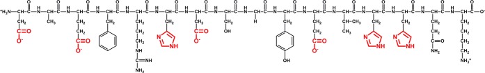
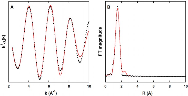
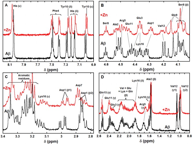
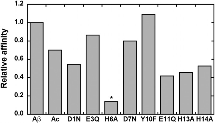
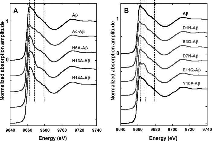
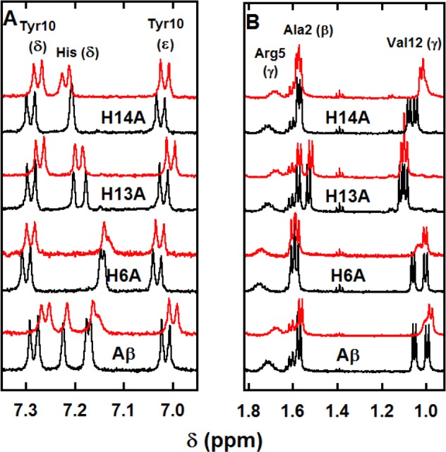
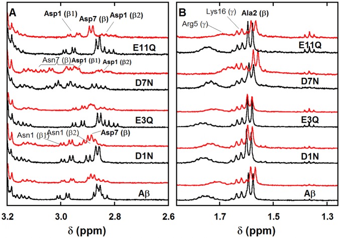
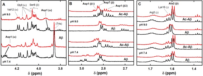
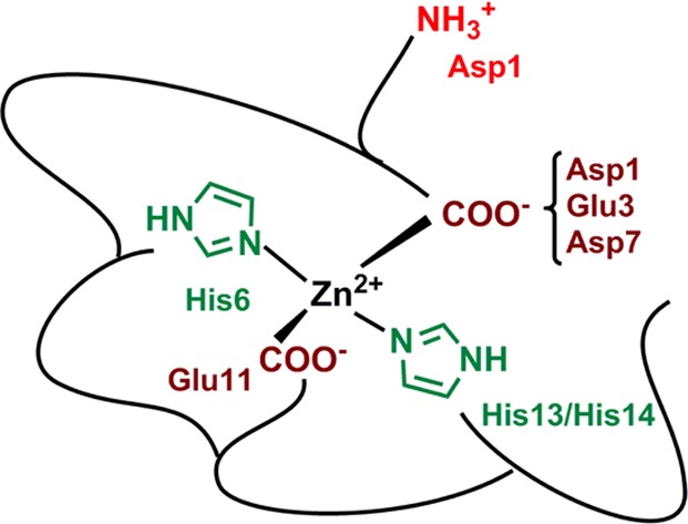
The Zn(II) ion has been linked to Alzheimer's disease (AD) due to its ability to modulate the aggregating properties of the amyloid-β (Aβ) peptide, where Aβ aggregation is a central event in the etiology of the disease. Delineating Zn(II) binding properties to Aβ is thus a prerequisite to better grasp its potential role in AD. Because of (i) the flexibility of the Aβ peptide, (ii) the multiplicity of anchoring sites, and (iii) the silent nature of the Zn(II) ion in most classical spectroscopies, this is a difficult task. To overcome these difficulties, we have investigated the impact of peptide alterations (mutations, N-terminal acetylation) on the Zn(Aβ) X-ray absorption spectroscopy fingerprint and on the Zn(II)-induced modifications of the Aβ peptides' NMR signatures. We propose a tetrahedrally bound Zn(II) ion, in which the coordination sphere is made by two His residues and two carboxylate side chains. Equilibria between equivalent ligands for one Zn(II) binding position have also been observed, the predominant site being made by the side chains of His6, His13 or His14, Glu11, and Asp1 or Glu3 or Asp7, with a slight preference for Asp1.


Conflict of interest statement

The authors declare no competing financial interest.

Figures

Scheme 1
Scheme 1. Aβ Peptide in the Protonation State Predominant at pH 7.4
The functional groups of the amino acid residues potentially involved in Zn(II) binding are highlighted in red.
Figure 1
Figure 1
k3-Weighted experimental (black dots) and least-squares fitted (red line) first coordination shell EXAFS spectra of the Zn(Aβ) at pH 6.9 (A) and the corresponding non-phase-shift-corrected Fourier transforms (B). Recording conditions: [Aβ] = 1.0 mM, [Zn(II)] = 0.9 mM in Hepes buffer 50 mM, T = 20 K.
Figure 2
Figure 2
1H NMR spectra of Aβ (bottom black lines) and of Aβ in the presence of 0.9 equiv of Zn(II) (top red lines) in selected regions (A: aromatic, B: Hα, C: Hβ, D: Hβ and Hγ, unless otherwise specified). [Aβ] = 300 μM, [Zn(II)] = 270 μM in d11-TRIS buffer 50 mM, pH = 7.4, T = 318 K, v = 500 MHz. δ(His6 Hδ) > δ(His14 Hδ) > δ(His13 Hδ). For the details of the amino acid residue nomenclature, see Scheme S1.
Figure 3
Figure 3
Zn(II) binding affinity values for the various modified peptides relative to Aβ. For the H6A mutant (*), the given value corresponds to a maximum value.
Figure 4
Figure 4
Zn(II) K-edge XANES spectra of Zn(II) bound to Aβ (black line) and to N mutants (panel A) and O mutants (panel B), Hepes buffer 50 mM pH 6.9, [Zn(II)] = 1.0 mM, [peptide] = 1.1 mM, T = 20 K. Normalization of the amplitude is given for the reference Zn(Aβ) complex.
Figure 5
Figure 5
1H NMR spectra of Aβ peptide and His-Ala mutants (bottom black lines) and of Aβ peptide and His-Ala mutants in the presence of 0.9 equiv of Zn(II) (top red lines) in selected regions (panel A: aromatic, panel B: Hβ and Hγ). [peptide] = 300 μM, [Zn(II)] = 270 μM in d11-TRIS buffer 50 mM, pH = 7.4, T = 318 K, v = 500 MHz.
Figure 6
Figure 6
1H NMR spectra of Aβ peptide and Asp-Asn and Glu-Gln mutants (bottom black lines) and of Aβ peptide and Asp-Asn and Glu-Gln mutants in the presence of 0.9 equiv of Zn(II) (top red lines) in selected regions (panel A: aromatic, panel B: Hβ and Hγ). [peptide] = 300 μM, [Zn(II)] = 270 μM in d11-TRIS buffer 50 mM, pH = 7.4, T = 318 K, v = 500 MHz.
Figure 7
Figure 7
1H NMR spectra of Aβ and Ac-Aβ peptides (bottom black lines) and of Aβ and Ac-Aβ peptides in the presence of 0.9 equiv of Zn(II) (top red lines) in selected regions as a function of pH (panel A: Hα, panel B: Asp Hβ, * stands for Asp7 protons, and panel C: Ala 2 Hβ). [peptide] = 300 μM, [Zn(II)] = 270 μM in d11-TRIS buffer 50 mM, pH = 7.4 or pH = 9.0, T = 318 K, v = 500 MHz.
Scheme 2
Scheme 2. Proposed Zn(II) Binding Site in Aβ (Predominant Species at pH 7.4)

References

    1. Holtzman D. M.; Morris J. C.; Goate A. M. Alzheimer’s disease: the challenge of the second century. Sci. Transl. Med. 2011, 3, 77sr1.10.1126/scitranslmed.3002369. - DOI - PMC - PubMed
    1. Kozlowski H.; Luczkowski M.; Remelli M.; Valensin D. Copper, Zinc and iron in neurodegenerative diseases (Alzheimer’s disease, Parkinson’s disease and prion diseases). Coord. Chem. Rev. 2012, 256, 2129–2141. 10.1016/j.ccr.2012.03.013. - DOI
    1. Hureau C. Coordination of redox active metal ions to the APP and to the amyloid-β peptides involved in AD. Part 1: an overview. Coord. Chem. Rev. 2012, 256, 2164–2174. 10.1016/j.ccr.2012.03.037. - DOI
    1. Noël S.; Cadet S.; Gras E.; Hureau C. The benzazole scaffold: a SWAT to combat Alzheimer’s Disease. Chem. Soc. Rev. 2013, 42, 7747–7762. 10.1039/c3cs60086f. - DOI - PubMed
    1. Viles J. H. Metal ions and amyloid formation in neurodegenerative diseases. Coord. Chem. Rev. 2012, 256, 2271–2284. 10.1016/j.ccr.2012.05.003. - DOI

MeSH terms

LinkOut - more resources