Review
. 2017 Feb:44:93-101.
doi: 10.1016/j.ceb.2016.09.003. Epub 2016 Sep 22.

Microtubule-organizing centers: from the centrosome to non-centrosomal sites

Affiliations
Review

Microtubule-organizing centers: from the centrosome to non-centrosomal sites

Ariana D Sanchez et al. Curr Opin Cell Biol. 2017 Feb.

Abstract

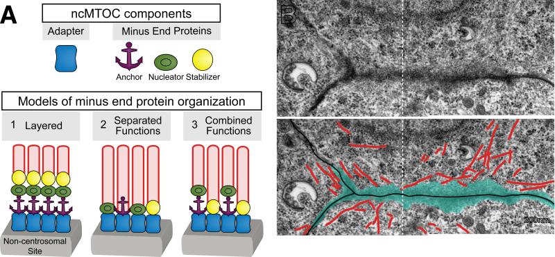
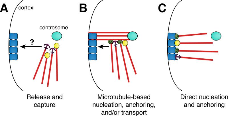
The process of cellular differentiation requires the distinct spatial organization of the microtubule cytoskeleton, the arrangement of which is specific to cell type. Microtubule patterning does not occur randomly, but is imparted by distinct subcellular sites called microtubule-organizing centers (MTOCs). Since the discovery of MTOCs fifty years ago, their study has largely focused on the centrosome. All animal cells use centrosomes as MTOCs during mitosis. However in many differentiated cells, MTOC function is reassigned to non-centrosomal sites to generate non-radial microtubule organization better suited for new cell functions, such as mechanical support or intracellular transport. Here, we review the current understanding of non-centrosomal MTOCs (ncMTOCs) and the mechanisms by which they form in differentiating animal cells.


Figures

Figure 1
Figure 1. Organization of MTOCs and microtubules in a variety of cell types
Microtubules (red) are organized by MTOCs (blue), the arrangement and localization of which varies with cell type. Drawings are not to scale.
Figure 2
Figure 2. ncMTOC structure and composition
(A) Cartoons depicting ncMTOC components and models for their arrangement at non-centrosomal sites. Cell-type specific adaptors (blue) bound to non-centrosomal sites (grey) interact with microtubule minus end proteins that anchor (purple), nucleate (green) and/or stabilize (yellow) microtubules (red). (1) Minus end proteins might be layered on top of adapters, which function together to sustain microtubules. (2) Alternatively, different minus end proteins could localize to independent minus ends, distributing their function between different populations of microtubules. (3) Finally, minus end proteins might colocalize at the same microtubule ends, functioning together to promote microtubule nucleation, anchoring, and/or stabilization. For example, NOCA-1 and γ-tubulin colocalize on microtubules in the C. elegans larval epidermis, but PTRN-1 does not and functions in a parallel pathway [25]. (B) An electron microscopy image of ncMTOCs (blue) at the apical membrane in C. elegans embryonic intestinal cells. Electron dense material (blue) is visible at the apical surfaces of three cells from which microtubules (red) emanate (partially reproduced from [54]). Note that two separate electron microscopy images have been overlaid (white dotted line).
Figure 3
Figure 3. Potential mechanisms for ncMTOC formation
(A) A division of labor model in which microtubules are nucleated at the centrosome, released, and then captured at a non-centrosomal site. Microtubules could be released with anchoring proteins attached or free minus ends could bind to anchoring and/or stabilizing proteins following their release. Microtubules are then transported to a non-centrosomal site via an unknown mechanism and captured by site-specific adapters. (B) Non-centrosomal microtubules could be nucleated, stabilized, and/or anchored from the sides of preexisting centrosomal microtubules and then transported along microtubules to non-centrosomal sites where they would interact with site specific adapters. (C) Microtubule minus end proteins could localize directly to non-centrosomal sites without a centrosome-based intermediate, where they would nucleate, stabilize, and anchor microtubules.

References

    1. Pickett-Heaps JD. The evolution of the mitotic apparatus: an attempt at comparative ultrastructural cytology in dividing plant cells. Cytobios. 1969;1:257–280.
    1. Porter KR. Cytoplasmic Microtubules and Their Functions. CIBA Foundation Symp. In: Wolstenholme GEW, O'Connor MJ, editors. Principles of Biomolecular Organizations. A. Churchill Ltd.; London: 1966. pp. 308–345.
    1. Voter WA, Erickson HP. he kinetics of microtubule assembly. Evidence for a two-stage nucleation mechanism. Journal of Biological Chemistry. 1984;259:10430–10438. - PubMed
    1. Gould RR, Borisy GG. The pericentriolar material in Chinese hamster ovary cells nucleates microtubule formation. The Journal of Cell Biology. 1977;73:601–615. - PMC - PubMed
    1. Woodruff JB, Wueseke O, Hyman AA. Pericentriolar material structure and dynamics. Phil. Trans. R. Soc. B. 2014;369:20130459–20130459. - PMC - PubMed

LinkOut - more resources