Suboptimal maternal diets alter mu opioid receptor and dopamine type 1 receptor binding but exert no effect on dopamine transporters in the offspring brain
- PMID: 27666382
- PMCID: PMC6288798
- DOI: 10.1016/j.ijdevneu.2016.09.008
Suboptimal maternal diets alter mu opioid receptor and dopamine type 1 receptor binding but exert no effect on dopamine transporters in the offspring brain
Abstract
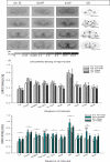
Birthweight is a marker for suboptimal fetal growth and development in utero. Offspring can be born large for gestational age (LGA), which is linked to maternal obesity or excessive gestational weight gain, as well as small for gestational age (SGA), arising from nutrient or calorie deficiency, placental dysfunction, or other maternal conditions (hypertension, infection). In humans, LGA and SGA babies are at an increased risk for certain neurodevelopmental disorders, including Attention Deficit/Hyperactivity Disorder, schizophrenia, and social and mood disorders. Using mouse models of LGA (maternal high fat (HF) diet) and SGA (maternal low protein (LP) diet) offspring, our lab has previously shown that these offspring display alterations in the expression of mesocorticolimbic genes that regulate dopamine and opioid function, thus indicating that these brain regions and neurotransmitter systems are vulnerable to gestational insults. Interestingly, these two maternal diets affected dopamine and opioid systems in somewhat opposing directions (e.g., LP offspring are generally hyperdopaminergic with reduced opioid expression, and the reverse is found for the HF offspring). These data largely involved evaluation at the transcriptional level, so the present experiment was designed to extend these analyses through an assessment of receptor binding. In this study, control, SGA and LGA offspring were generated from dams fed control, low protein or high fat diet, respectively, throughout pregnancy and lactation. At weaning, mice were placed on the control diet and sacrificed at 12 weeks of age. In vitro autoradiography was used to measure mu-opioid receptor (MOR), dopamine type 1 receptor (D1R), and dopamine transporter (DAT) binding level in mesolimbic brain regions. Results showed that the LP offspring (males and females) had significantly higher MOR and D1R binding than the control animals in the regions associated with reward. In HF offspring there were no differences in MOR binding, and limited increases in D1R binding, seen only in females in the nucleus accumbens core and the dorsomedial caudate/putamen. DAT binding revealed no differences in either models. In conclusion, LP but not HF offspring show significantly elevated MOR and D1R binding in the brain thus affecting DA and opioid signaling. These findings advance the current understanding of how suboptimal gestational diets can adversely impact neurodevelopment and increase the risk for disorders such as ADHD, obesity and addiction.
Keywords: Dopamine; Maternal high fat diet; Maternal low protein diet; Opioid; Reward.
Copyright © 2016 ISDN. Published by Elsevier Ltd. All rights reserved.
Figures


References
-
- Barker DJP. In utero programming of chronic disease. Clinical Science. 1998;95:115–128. - PubMed
-
- Barker DJP, Osmond C. Infant mortality, childhood nutrition, and ischaemic heart disease in England and Wales. Lancet. 1986;1:1077–1081. - PubMed
-
- Bergevin A, Girardot Daphne, Bourque Marie-Josee, Trudeau L-E. Presynaptic μ-opioid receptors regulate a late step of the secretory process in rat ventral tegmental area GABAergic neurons. Neuropharmacology. 2002;42:1065–1078. - PubMed
-
- Brawarsky P, Stotland NE, Jackson RA, Fuentes-Afflick E, Escobar GJ, Rubashkin N, Haas JS. Pre-pregnancy and pregnancy-related factors and the risk of excessive or inadequate gestational weight gain. International Journal of Gynecology & Obstetrics. 2005b;91:125–131. - PubMed
MeSH terms
Substances
Grants and funding
LinkOut - more resources
Full Text Sources
Other Literature Sources
Research Materials
Miscellaneous

