. 2017 Jan:45:64-68.e5.
doi: 10.1016/j.exphem.2016.09.006. Epub 2016 Sep 22.

The AMP-activated protein kinase beta 1 subunit modulates erythrocyte integrity

Affiliations

The AMP-activated protein kinase beta 1 subunit modulates erythrocyte integrity

Emma L Cambridge et al. Exp Hematol. 2017 Jan.

Abstract

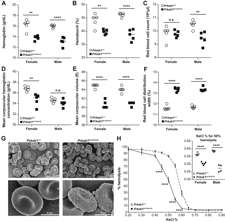
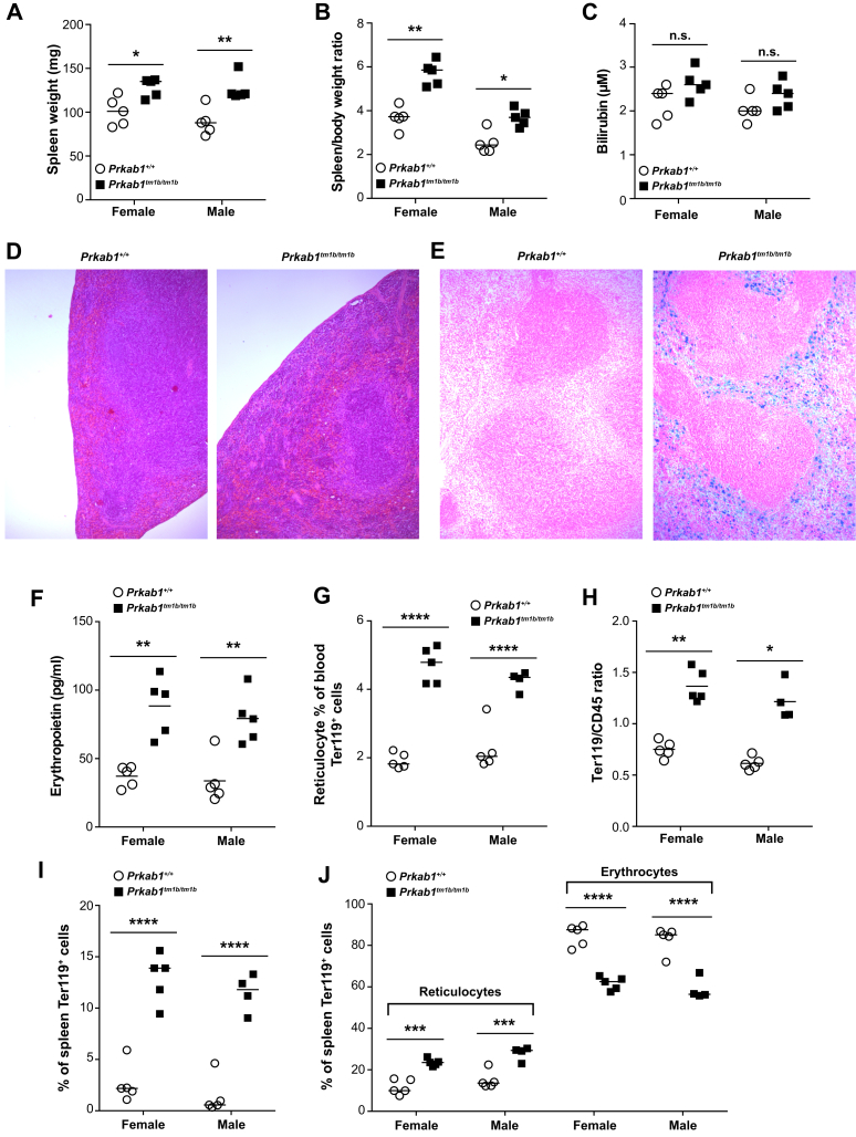
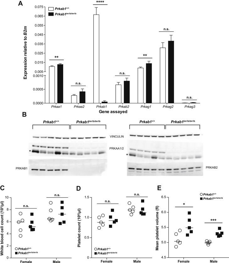
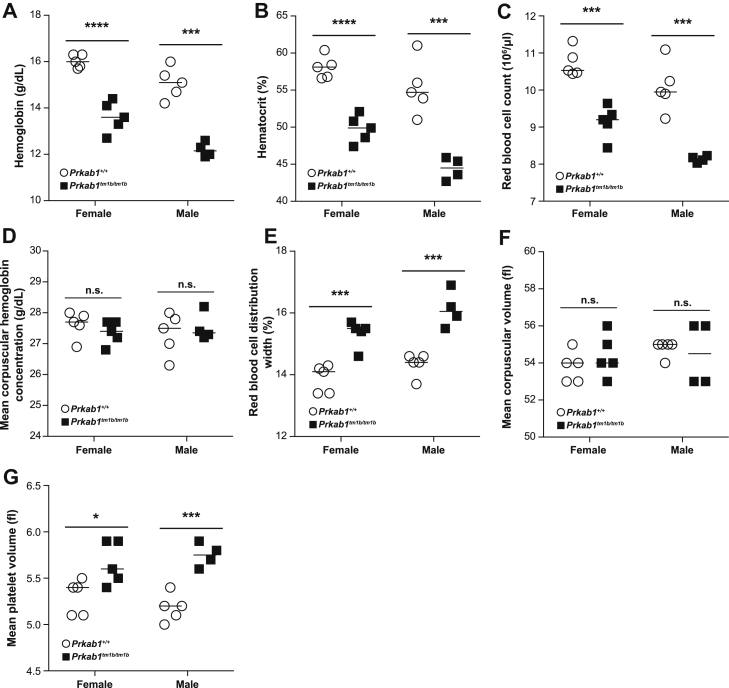
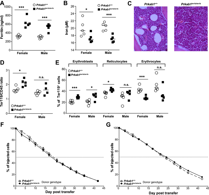
Failure to maintain a normal in vivo erythrocyte half-life results in the development of hemolytic anemia. Half-life is affected by numerous factors, including energy balance, electrolyte gradients, reactive oxygen species, and membrane plasticity. The heterotrimeric AMP-activated protein kinase (AMPK) is an evolutionarily conserved serine/threonine kinase that acts as a critical regulator of cellular energy balance. Previous roles for the alpha 1 and gamma 1 subunits in the control of erythrocyte survival have been reported. In the work described here, we studied the role of the beta 1 subunit in erythrocytes and observed microcytic anemia with compensatory extramedullary hematopoiesis together with splenomegaly and increased osmotic resistance.


Figures

Figure 1
Figure 1
Prkab1-deficient mice present with anemia, erythrocyte morphologic abnormalities, and increased erythrocyte osmotic resistance. (A) Hemoglobin, (B) hematocrit, (C) red blood cell count, (D) mean corpuscular hemoglobin concentration, (E) mean corpuscular volume, and (F) red blood cell distribution width of 16-week-old Prkab1+/+ and Prkab1tm1b/tm1b mice. **p < 0.01, and ****p < 0.0001, unpaired two-tailed Student t test. (G) Representative SEM images of erythrocytes from Prkab1+/+ and Prkab1tm1b/tm1b mice. (H) Osmotic resistance of Prkab1+/+ and Prkab1tm1b/tm1b erythrocytes (combined males and females). ****p < 0.0001 as determined by a repeated-measures two-way analysis of variance with Sidak's multiple comparison test adjusting for multiple testing, the insert is % of NaCl for 50% hemolysis of erythrocytes ****p < 0.0001, unpaired two-tailed Student t test. All data are representative of three independent experiments or two mice for SEM analysis. Each symbol represents an individual mouse with the line at the mean except for (H), where n = 10 for Prkab1+/+ and n = 9 for Prkab1tm1b/tm1b with mean ± standard error of the mean.
Figure 2
Figure 2
Prkab1-deficient mice have splenomegaly, extramedullary hematopoiesis, and splenic iron deposits. (A) Spleen weight. (B) Spleen/body weight ratio (mg/g). (C) Plasma bilirubin concentration. (D) Hematoxylin and eosin-stained sections of spleen (100× magnification). (E) Perls’-stained sections of spleen (100× magnification). (F) Plasma erythropoietin. (G) Percentage of circulating reticulocytes. (H) Splenic erythroid (Ter119)/leukocyte (CD45) ratio. (I) Percentage of splenic erythroblasts. (J) Percentages of splenic reticulocytes and erythrocytes. For all, *p < 0.05, **p < 0.01, ***p < 0.001, and ****p < 0.0001, unpaired two-tailed Student t test except for spleen/body weight ratio and Ter119/CD45 ratio, which were analyzed with a Mann–Whitney test. All data are representative of three independent experiments or four mice for histology analysis; each symbol represents an individual mouse with the line at the mean.
Supplementary Figure E1
Molecular and phenotypic characterization of Prkab1-deficient mice. (A) Mean gene expression of AMPK subunits in Prkab1+/+ and Prkab1tm1b/tm1b spleen RNA. n = 5 per genotype, with error bars representing standard errors of the mean. (B) Immunoblot analysis of AMPK subunits in Prkab1+/+ and Prkab1tm1b/tm1b spleen protein lysates. *IgG heavy chain. (C) White blood cell count. (D) Mean platelet volume. (E) Platelet count of 16-week-old Prkab1+/+ and Prkab1tm1b/tm1b mice. *p < 0.05, **p < 0.01, *** p < 0.001, and ****p < 0.0001. Student’s t test, hematology data are representative of three independent experiments. Each symbol represents an individual mouse with the line at the mean.
Supplementary Figure E2
Prkab1tm1b/tm1b mice have altered hematologic parameters at 4 weeks of age. (A) Hemoglobin, (B) hematocrit, (C) red blood cell count, (D) mean corpuscular hemoglobin concentration, (E) red blood cell distribution width, (F) mean corpuscular volume, and (G) mean platelet volume of 4-week-old Prkab1+/+ and Prkab1tm1b/tm1b mice. *p < 0.05, ***p < 0.001, and ****p < 0.0001, unpaired two-tailedStudent t test. Data are representative of two independent experiments. Each symbol represents an individual mouse with the line at the mean.
Supplementary Figure E3
Characterofization of circulating iron, bone marrow erythropoiesis, and erythrocyte half-life of Prkab1-deficient mice. (A) Plasma ferritin concentration. (B) Plasma iron concentration. (C) Representative H&E stained bone marrow sections from Prkab1+/+ and Prkab1tm1b/tm1b mice (400× magnification). (D) Erythroid (Ter119)/Leukocyte (CD45) ratio of bone marrow from Prkab1+/+ and Prkab1tm1b/tm1b mice. (E) Characterization of erythropoiesis in the bone marrow of Prkab1+/+ and Prkab1tm1b/tm1b mice. In vivo half-life of erythrocytes transferred into Prkab1+/+ (F) or Prkab1tm1b/tm1b (G) mice. For all, *p < 0.05, ***p < 0.001, and ****p < 0.0001, unpaired two-tailed Student t test, except for Ter119/CD45 ratio, which was analyzed with a Mann–Whitney test. Data are representative of two independent experiments or four mice for histology analysis. Each symbol represents an individual mouse with the line at the mean, except for (F) and (G), where n = 5 with mean ± standard error of the mean.

References

    1. Mohandas N., Gallagher P.G. Red cell membrane: Past, present, and future. Blood. 2008;112:3939–3948. - PMC - PubMed
    1. Carling D., Thornton C., Woods A., Sanders M.J. AMP-activated protein kinase: New regulation, new roles? Biochem J. 2012;445:11–27. - PubMed
    1. Hardie D.G. AMP-activated/SNF1 protein kinases: Conserved guardians of cellular energy. Nat Rev Mol Cell Biol. 2007;8:774–785. - PubMed
    1. Foretz M., Hebrard S., Guihard S. The AMPK gamma1 subunit plays an essential role in erythrocyte membrane elasticity, and its genetic inactivation induces splenomegaly and anemia. FASEB J. 2011;25:337–347. - PubMed
    1. Wang S., Dale G.L., Song P., Viollet B., Zou M.H. AMPKalpha1 deletion shortens erythrocyte life span in mice: Role of oxidative stress. J Biol Chem. 2010;285:19976–19985. - PMC - PubMed

LinkOut - more resources