. 2016 Oct 5;92(1):100-113.
doi: 10.1016/j.neuron.2016.08.038. Epub 2016 Sep 22.

How Azobenzene Photoswitches Restore Visual Responses to the Blind Retina

Affiliations

How Azobenzene Photoswitches Restore Visual Responses to the Blind Retina

Ivan Tochitsky et al. Neuron. .

Abstract

Azobenzene photoswitches confer light sensitivity onto retinal ganglion cells (RGCs) in blind mice, making these compounds promising candidates as vision-restoring drugs in humans with degenerative blindness. Remarkably, photosensitization manifests only in animals with photoreceptor degeneration and is absent from those with intact rods and cones. Here we show that P2X receptors mediate the entry of photoswitches into RGCs, where they associate with voltage-gated ion channels, enabling light to control action-potential firing. All charged photoswitch compounds require permeation through P2X receptors, whose gene expression is upregulated in the blind retina. Photoswitches and membrane-impermeant fluorescent dyes likewise penetrate through P2X receptors to label a subset of RGCs in the degenerated retina. Electrophysiological recordings and mapping of fluorescently labeled RGC dendritic projections together indicate that photosensitization is highly selective for OFF-RGCs. Hence, P2X receptors are a natural conduit allowing cell-type-selective and degeneration-specific delivery of photoswitches to restore visual function in blinding disease.

Keywords: P2X receptor; azobenzene; blindness; ion channel; photoswitch; retina; retinal degeneration; retinal ganglion cell; retinitis pigmentosa; vision.


Figures

Figure 1
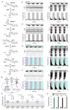
Figure 1. Photoswitch compounds photosensitize RGCs from rd1 but not WT mice
A) Photoswitch structures. Visible light converts DENAQ and BENAQ from trans to cis; the compounds then quickly relax back to trans in the dark. 380nm light converts AAQ and QAQ from trans to cis and then the compounds can be converted back to trans by 500nm light. B) Ion channels targeted by the photoswitches. DENAQ, BENAQ and AAQ block HCN channels and K+ channels, while QAQ blocks Na+ and K+ channels but not HCN channels. C–F) MEA recordings of RGC activity from synaptically isolated WT (left) and rd1 (right) mouse RGCs treated with DENAQ (C), BENAQ (D), AAQ (E) or QAQ (F). Light stimuli (top), raster plots of RGC activity (middle), average firing rate plots (bottom). G) PI value box plots - median (red), 1st/3rd quartiles (blue) with outliers (green) for RGCs from photoswitch treated WT and rd1 mouse retinas. Ns are RGCs. P-values obtained using rank sum test. H) Average PI values from photoswitch treated synaptically isolated WT and rd1 mouse retinas. Data are mean±SEM. See also Figure S1, Table S2.
Figure 2
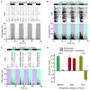
Figure 2. Photoswitches enter rd1 RGCs through activated P2X receptors
A) Representative MEA recording from rd1 RGCs treated with DENAQ and the P2X receptor antagonist TNP-ATP. Photoswitching is absent. B) PI values from in vitro photoswitch treated rd1 retinas after pretreatment with P2X and TRPV1 receptor antagonists. C) PI values from in vitro DENAQ treated rd1 retinas after pretreatment with apyrase with or without the addition of ATPγS. D) PI values from rd1 retinas treated in vitro with a photoswitch alone or with bzATP. P values in panels B–D compare PI values for rd1 retinas treated with photoswitch alone vs photoswitch + specified compound(s). All data are mean±SEM. See also Figures S2, S3, Table S2.
Figure 3
Figure 3. HCN channels are necessary for RGC photosensitization by some, but not all, photoswitches
A–C) MEA recordings from synaptically isolated rd1 RGCs treated with BENAQ (A), QAQ (B) or AAQ (C) and the HCN channel blocker ivabradine. BENAQ-treated RGCs (A) are no longer light sensitive after HCN channel block. Both QAQ-treated (B) and AAQ-treated (C) RGCs remain photosensitive even after HCN channel block, but the polarity of photoswitching is reversed in AAQ-treated RGCs (C). D) PI values from rd1 retinas treated with various photoswitches alone or with photoswitches followed by ivabradine. Data are mean±SEM. See also Table S2.
Figure 4
Figure 4. Genes encoding photoswitch conduits and targets are up-regulated in WT and rd1 mouse retinas
A–C) Differences in gene expression in rd1 retinas relative to WT retinas based on the ddCt values from RT-qPCR, normalized to β-actin (Actb) expression. Data are mean±SEM, n=5 retinas. See also Figure S4, Table S1.
Figure 5
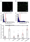
Figure 5. P2X receptors are necessary for dye entry into RGCs in the rd1 retina
A) WT retina loading of YO-PRO-1 (green) shows loading primarily in vasculature with neuronal populations being almost completely absent of YO-PRO-1 labeling. B) By contrast, a subpopulation of rd1 RGCs are readily labeled with YO-PRO-1. C–D) Fluorescence histograms of YO-PRO-1 labeling. The threshold for labeled cells was set at +2SD above autofluorescence values. YO-PRO-1 labels a larger fraction of rd1 RGCs (C) compared to WT RGCs (D) (p<0.001). E) Percentage of YO-PRO labeled cells in rd1 and WT RGCs. YO-PRO labeled WT RGCs=3.7±1.3%, n=1854; rd1=29.0±6.9%, n=1106, p<0.001, two-tailed unpaired Student’s t-test. P2X channel blockers TNP-ATP (5±2%) and A740003 (9±1.5%); and breakdown of extracellular ATP with apyrase (12±5%) all reduced YO-PRO labeling while capsazepine had no effect. Data are mean±SEM. See also Figure S5.
Figure 6
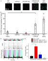
Figure 6. Activation of over-expressed P2X7R enables permeation of dye and photoswitch into WT RGCs
A–B) Confocal images (A) and quantification (B) of YO-PRO-1 loading (green) into WT RGCs. WT RGCs are not labeled with YO-PRO after addition of exogenous ATPγS or P2X7R overexpression (OE) alone. P2X7R OE together with the addition of ATPγS enable dye loading into WT RGCs (2×2 ANOVA p<0.0001, TukeyHSD Interaction p<0.01). By comparison, YO-PRO-1 robustly loads into rd1 RGCs without any genetic or pharmacological manipulation. C) Sample MEA recording from synaptically isolated WT RGCs overexpressing P2X7 and treated with QAQ+bzATP. D) PI values from photoswitch treated WT mouse retinas overexpressing P2X7 and treated with bzATP. Data are mean±SEM. See also Figure S6, Table S2.
Figure 7
Figure 7. P2X receptor permeant dyes selectively enter OFF-RGCs in rd1 retina
A) (Top) Rd1 RGC filled with a cytoplasmic fluorophore (Alexa 488). The outlined area was aggregated to a single plane for analysis (Bottom) Nuclear-ID labeling of RGCs (red). B) Orthogonal views of aggregated fluorescence data show the GCL and INL. The location of the peak of the dendritic fluorescence was used to differentiate between putative ON and OFF RGCs. C) RGCs with dendrites stratifying in the ON sublamina lacked the nuclear labeling that is present in those cells whose dendrites lay in the OFF sublamina. D) RGC YO-PRO nuclear fluorescence plotted as function of RGC dendritic stratification in the IPL. Cells with nuclear labeling index greater than 1 (red dashed line) are above threshold for labeling. Cells with dendrites deeper in the IPL (above black dashed line) are readily labeled by YO-PRO. E) Nuclear labeling index for putative ON (n=17) and OFF (n=12) RGCs as well as bistratified ON/OFF RGCs (n=9) shows preferential loading in ON RGCs. Data are mean±SEM. (1×3 ANOVA p<0.001, TukeyHSD p<0.001)
Figure 8
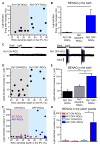
Figure 8. BENAQ selectively photosensitizes OFF-RGCs in rd1 retina
A) Light-elicited increase in firing rate in BENAQ-treated rd1 RGCs plotted in relation to the depth of the RGC dendritic stratification in the IPL. Bistratified ON/OFF RGCs are not shown. Putative ON-RGC/OFF-RGC classification shown in gray/blue. B) Light elicits a larger firing rate increase in BENAQ-treated OFF-RGCs (mean=6.4 Hz, n=6) compared to ON-RGCs (0.2 Hz, n=7, p=0.016). Light evoked no response in ON-OFF RGCs (n=6). C) Example traces from BENAQ-treated ON (left) and OFF (right) RGCs. Bar above the trace represents the presence or absence of light. D) Light elicited increase in Ih in extracellular bath BENAQ-treated rd1 RGCs plotted in relation to the depth of the RGC dendritic stratification in the IPL. Putative ON/OFF-RGC classification shown in gray/blue. E) Average light elicited increase in Ih in extracellular bath BENAQ-treated OFF-RGCs (mean=151.8±15.5 pA, n=5), ON/OFF-RGCs (mean=80.6±9.3 pA, n=6, p<0.001), and ON-RGCs (mean=45.3±10.7 pA, n=11, p<0.001, Bonferroni post hoc test). Data are mean±SEM. F) Light elicited increase in Ih in intracellular patch-pipette BENAQ-treated WT (red) and rd1 (blue) RGCs plotted in relation to the depth of the RGC dendritic stratification in the IPL. G) Average light elicited increase in Ih in intracellular patch-pipette BENAQ-treated WT ON-RGCs (mean=7.8±5.6 pA, n=5), WT OFF-RGCs (mean=3.9±1.0 pA, n=5), rd1 ON-RGCs (mean=7.0±5.4 pA, n=6), and rd1 ON-RGCs (mean=135±65 pA, n=8, p=0.046). Data are mean±SEM. See also Figure S7.

References

    1. Banghart MR, Mourot A, Fortin DL, Yao JZ, Kramer RH, Trauner D. Photochromic blockers of voltage-gated potassium channels. Angewandte Chemie. 2009;48:9097–9101. - PMC - PubMed
    1. Barboni MTS, Nagy BV, de Araújo Moura AL, Damico FM, da Costa MF, Kremers J, Ventura DF. ON and OFF Electroretinography and Contrast Sensitivity in Duchenne Muscular Dystrophy. Investigative ophthalmology & visual science. 2013;54:3195–3204. - PubMed
    1. Bemme S, Weick M, Gollisch T. Pharmacological blockade of HCN channels changes evoked and spontaneous activity of retinal ganglion cells. Investigative ophthalmology & visual science. 2014;55:2385–2385.
    1. Brandle U, Guenther E, Irrle C, Wheeler-Schilling TH. Gene expression of the P2X receptors in the rat retina. Brain research Molecular brain research. 1998;59:269–272. - PubMed
    1. Browne LE, Compan V, Bragg L, North RA. P2X7 receptor channels allow direct permeation of nanometer-sized dyes. The Journal of neuroscience : the official journal of the Society for Neuroscience. 2013;33:3557–3566. - PMC - PubMed

MeSH terms