. 2016 Sep 26:6:34072.
doi: 10.1038/srep34072.

In vitro and in vivo study of additive manufactured porous Ti6Al4V scaffolds for repairing bone defects

Affiliations

In vitro and in vivo study of additive manufactured porous Ti6Al4V scaffolds for repairing bone defects

Guoyuan Li et al. Sci Rep. .

Abstract

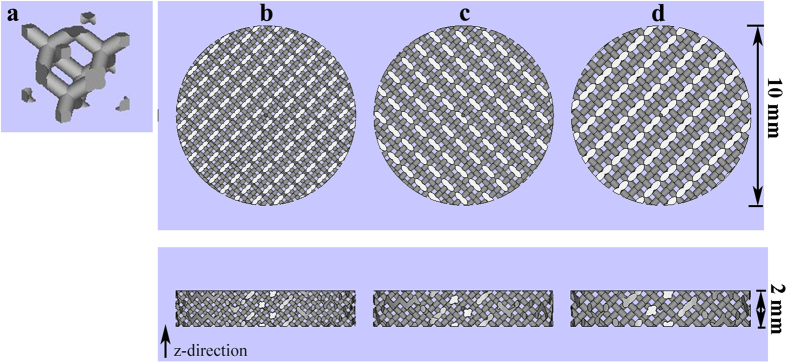
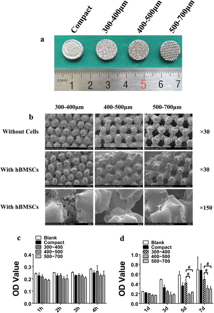
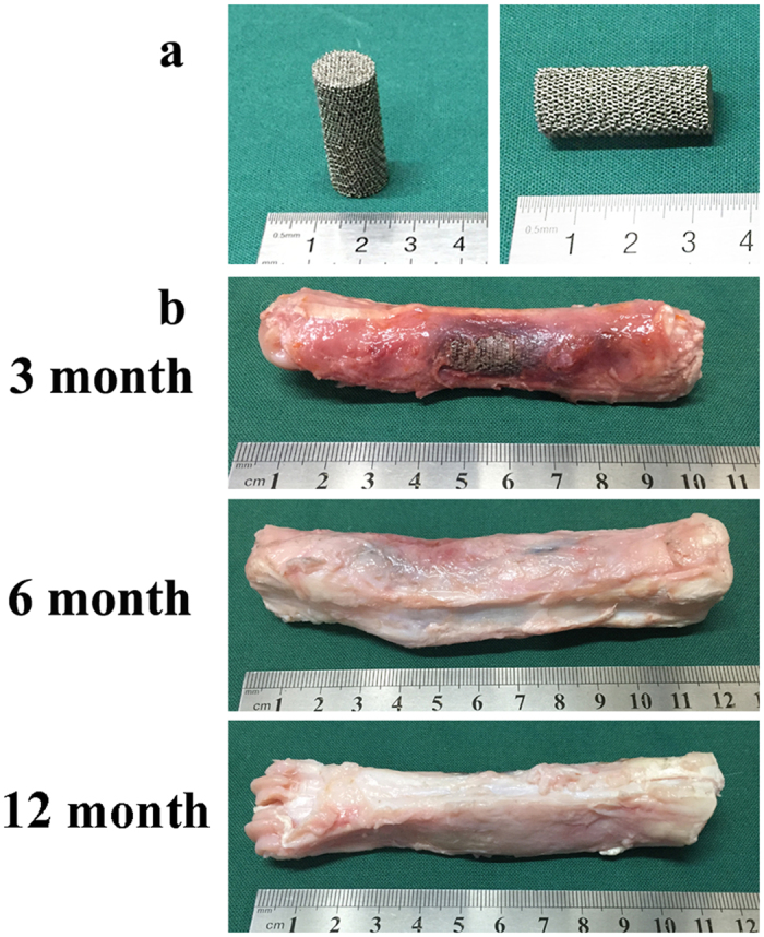
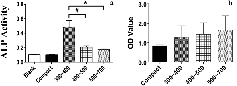
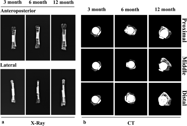
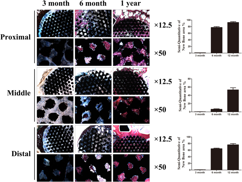
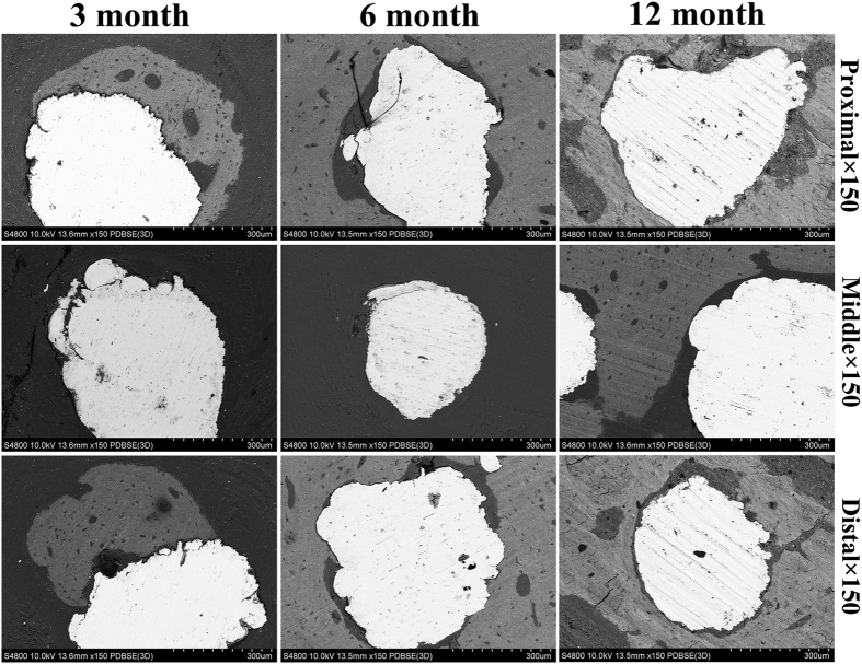
Metallic implants with a low effective modulus can provide early load-bearing and reduce stress shielding, which is favorable for increasing in vivo life-span. In this research, porous Ti6Al4V scaffolds with three pore sizes (300~400, 400~500, and 500~700 μm) were manufactured by Electron Beam Melting, with an elastic modulus range of 3.7 to 1.7 GPa. Cytocompatibility in vitro and osseointegration ability in vivo of scaffolds were assessed. hBMSCs numbers increased on all porous scaffolds over time. The group with intended pore sizes of 300 to 400 μm was significantly higher than that of the other two porous scaffolds at days 5 and 7. This group also had higher ALP activity at day 7 in osteogenic differentiation experiment. The scaffold with pore size of 300 to 400 μm was implanted into a 30-mm segmental defect of goat metatarsus. In vivo evaluations indicated that the depth of bone ingrowth increased over time and no implant dislocation occurred during the experiment. Based on its better cytocompatibility and favorable bone ingrowth, the present data showed the capability of the additive manufactured porous Ti6Al4V scaffold with an intended pore size of 300 to 400 μm for large segmental bone defects.


Figures

Figure 1
Figure 1. Computer aid design of porous Ti6Al4V alloy scaffolds with different pore size used in vitro.
(a) A single unit of diamond-shaped lattice; (b) pore size of 300~400 μm; (c) pore size of 400~500 μm; (d) pore size of 500~700 μm.
Figure 2
Figure 2. Scaffold appearance and cell adhesion, growth and morphology on the tested scaffolds.
(a) Scaffolds used in the in vitro experiment with a height and diameter of 2 and 10 mm, respectively. (b) SEM micrographs of porous Ti6Al4V scaffolds and cell morphology on porous scaffolds after being cultured for 7 days. The cells were mainly located between the gaps of the struts, flattened and spread well with numerous filopodia extensions, and the cell number in the group of intended pore size of 300 to 400 μm was higher than that of the other two groups. (c) Cell adhesion on the tested scaffolds. A higher OD value indicates that more cells adhered on the scaffolds. (d) Cell growth on the tested scaffolds. A higher OD value indicates that more cells grew or remained “alive” on the scaffolds (#p < 0.05).
Figure 3
Figure 3
Scaffolds used in the in vivo experiment with a height and diameter of 30 and 10 mm (a) gross specimens at different time points (3 months, 6 months and 12 months) (b).
Figure 4
Figure 4. Cell osteogenic differentiation in vitro.
ALP activity at day 7 (a) and semi-quantitative analysis of calcium nodule at day 21 (b) on porous Ti6Al4V scaffolds and the control samples. A high OD value indicates that much more calcium nodule formed on the scaffolds (#p < 0.05, *p < 0.01).
Figure 5
Figure 5
Plain radiographs (anterior-posterior and lateral) (a) and CT scans of specimens (proximal, middle and distal positions) (b) at different time points (3 months, 6 months and 12 months). New bone formation appeared on the lateral side opposite to the plate, and continuous mature bone was observed around the scaffolds at 1 year.
Figure 6
Figure 6
Histologic sections of porous Ti6Al4V scaffolds implanted into goat metatarsus large segmental defects and semi-quantitative of new bone area in the scaffolds. At 3 months, callus in the periphery regions had formed; apparent bone ingrowth was observed at 6 months; at 12 months, the inner space of the scaffolds was nearly completely filled with bone tissue. New bone area increased with time and, compared with the middle position, both ends had relatively more amounts of new bone, with the proximal position being superior to the distal position. Stain: Stevenel’s blue and Van Gieson’s picrofuchsin. Purple indicates bone; black indicates materials; blue indicates fibrovascular tissue.
Figure 7
Figure 7. SEM micrographs of bone apposition and bone microstructure on porous scaffolds in different positons at 3 month, 6month and 12 month.
White indicate Ti6Al4V, grey indicate new bone.

References

    1. De Long W. G. Jr. et al. Bone grafts and bone graft substitutes in orthopaedic trauma surgery. A critical analysis. J. Bone Joint Surg. Am. 89, 649–658 (2007). - PubMed
    1. Kurien T., Pearson R. G. & Scammell B. E. Bone graft substitutes currently available in orthopaedic practice: the evidence for their use. Bone Joint J. 95B, 583–597 (2013). - PubMed
    1. LeGeros R. Z. Properties of osteoconductive biomaterials: calcium phosphates. Clin. Orthop. Relat. Res. 395, 81–98 (2002). - PubMed
    1. Brånemark P. I. et al. Osseointegrated implants in the treatment of the edentulous jaw. Experience from a 10-year period. Scand. J. Plast Reconstr. Surg. Suppl. 16, 132 (1977). - PubMed
    1. Khan S. N., Tomin E. & Lane J. M. Clinical applications of bone graft substitutes. Orthop. Clin. North Am. 31, 389–398 (2000). - PubMed