. 2016 Sep 26:7:12837.
doi: 10.1038/ncomms12837.

The structural basis for CD36 binding by the malaria parasite

Affiliations

The structural basis for CD36 binding by the malaria parasite

Fu-Lien Hsieh et al. Nat Commun. .

Abstract

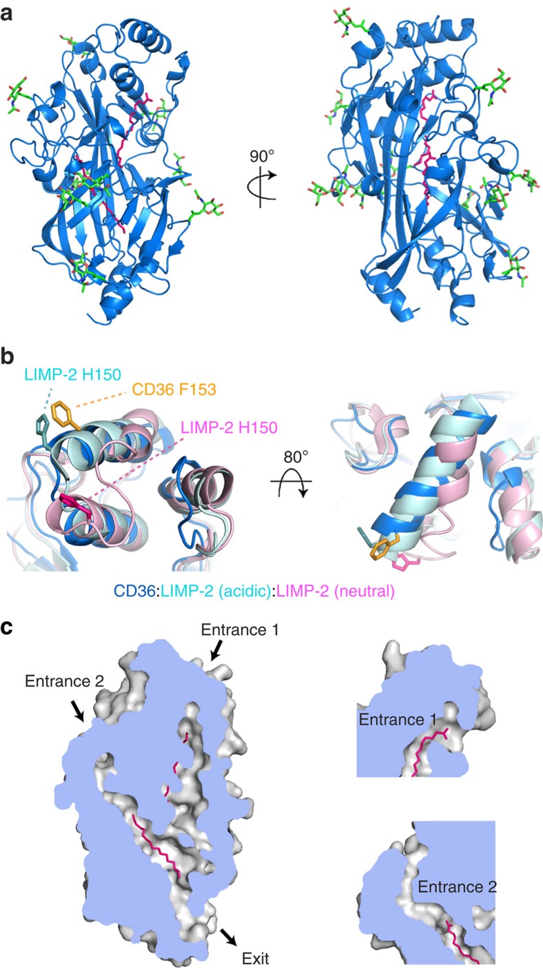
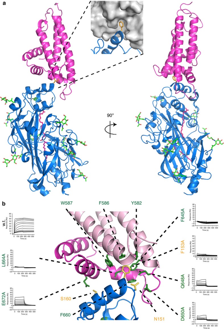
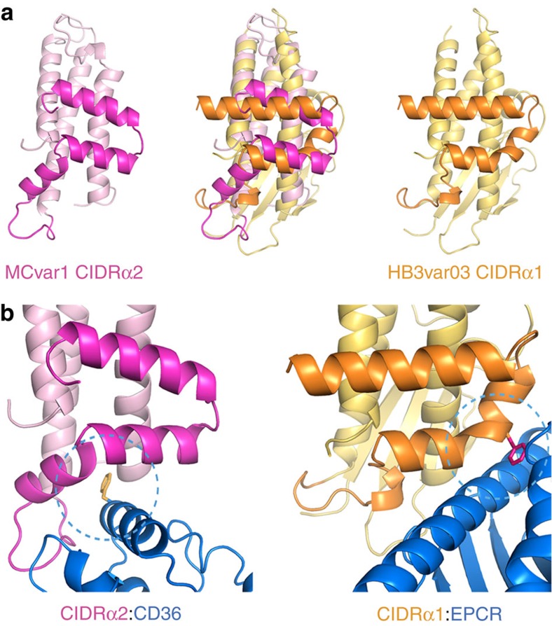
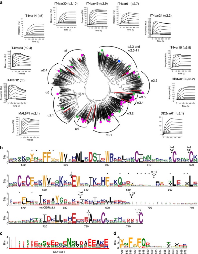
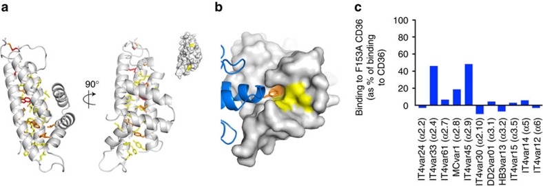
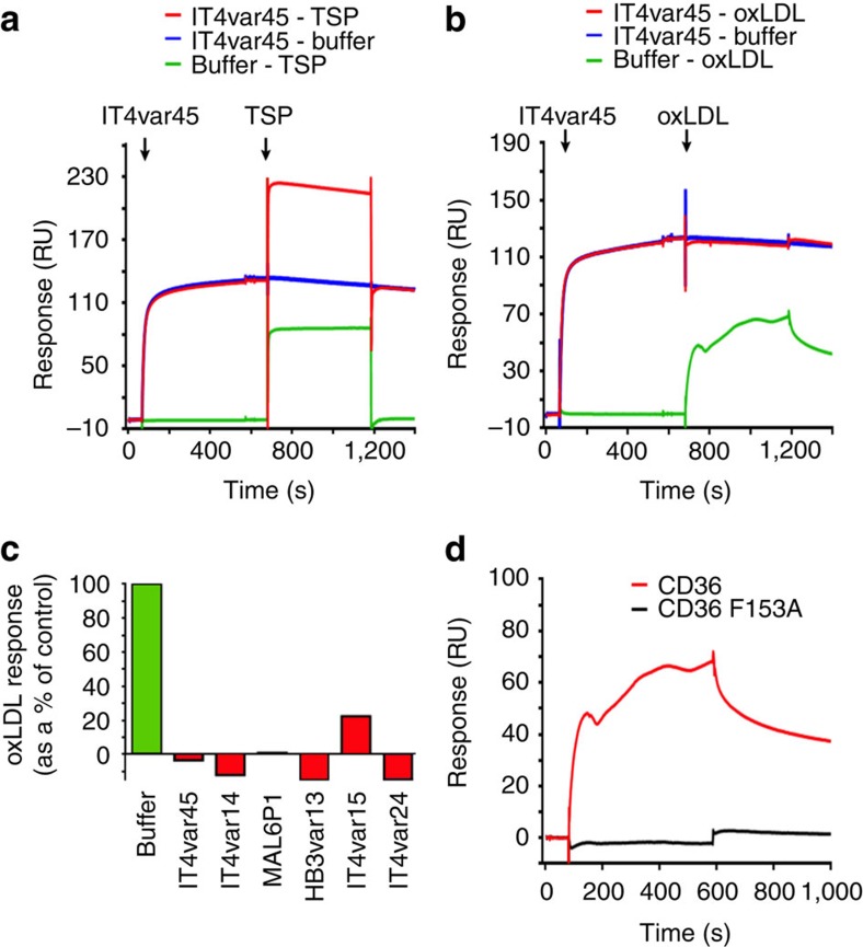
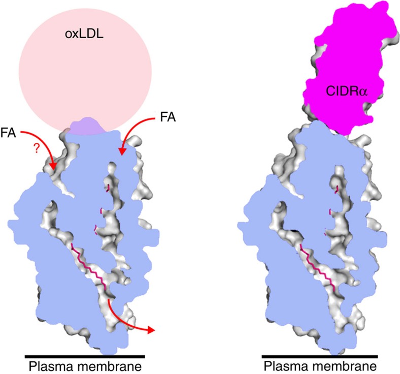
CD36 is a scavenger receptor involved in fatty acid metabolism, innate immunity and angiogenesis. It interacts with lipoprotein particles and facilitates uptake of long chain fatty acids. It is also the most common target of the PfEMP1 proteins of the malaria parasite, Plasmodium falciparum, tethering parasite-infected erythrocytes to endothelial receptors. This prevents their destruction by splenic clearance and allows increased parasitaemia. Here we describe the structure of CD36 in complex with long chain fatty acids and a CD36-binding PfEMP1 protein domain. A conserved hydrophobic pocket allows the hugely diverse PfEMP1 protein family to bind to a conserved phenylalanine residue at the membrane distal tip of CD36. This phenylalanine is also required for CD36 to interact with lipoprotein particles. By targeting a site on CD36 that is required for its physiological function, PfEMP1 proteins maintain the ability to tether to the endothelium and avoid splenic clearance.


Figures

Figure 1
Figure 1. The structure of CD36 and its binding of fatty acids.
(a) The structure of CD36, shown in blue. The nine N-linked glycosylation sites and associated sugars are green while two palmitic acids are shown as pink sticks. (b) An alignment of CD36 (blue) with the structures of LIMP-2 at acidic (cyan) and neutral (pink) pH. Residues F153 from CD36 (orange) and H150 from LIMP-2 at acidic (light blue) and neutral (pink) pH are highlighted. (c) A section through a surface representation of CD36 showing the central core cavity occupied by two palmitic acids (pink). Insets show two putative entrances to this central cavity.
Figure 2
Figure 2. The structural basis for CD36 binding by PfEMP1 proteins.
(a) The structure of the complex of CD36 (blue) with the CIDRα2.8 domain of the MCvar1 PfEMP1 protein (pink). N-linked glycans are shown in green. The inset shows a surface representation of the CIDRα domain with CD36 as a blue cartoon and F153 of CD36 in orange. (b) A close-up view of the interface between CD36 and the CIDRα domain with the key interacting residues labelled. The three-core α-helices of the CIDRα domain (α1, α2 and α6) are coloured in light pink while the insert (α3-α5) is dark pink. Also shown is the effect of mutagenesis of key interacting residues as determined by surface plasmon resonance. The most critical interactions are mediated by F153 of CD36, which fits into a hydrophobic pocket lined by residues including F645 and L664 of the CIDRα domain.
Figure 3
Figure 3. A comparison of the features that allow CIDRα domains to bind to EPCR or CD36.
(a) A comparison of the structure of a CD36-binding CIDRα2 domain (pink) with that of an EPCR-binding CIDRα1 domain (orange). Both domains share a core three α-helical bundle. The insertion that emerges between the second and third of these core helices forms a docking platform for ligands. (b) A close up of the binding interfaces that mediates the CIDRα2:CD36 and CIDRα1:EPCR interactions. The CIDRα1 domains have a phenylalanine residue (F656) on a convex surface of the domain that protrudes into the hydrophobic groove of EPCR. In contrast, the CIDRα2 domains have a hydrophobic pocket that binds to a protruding phenyalanine residue (F153) from CD36.
Figure 4
Figure 4. Extensive diversity in CD36 binding CIDRα domains.
(a) Sequence distance tree of 2386 full-length CIDRα2-6 domains. Red lines represent sequences from Plasmodium reichenowi. A blue circle marks the sequence of the crystallized CIDRα2.8 domain (also see Supplementary Fig. 8). Green circles mark sequences of recombinant CIDRα domains for which the affinity for CD36 binding were tested (corresponding SPR traces given). Pink circles mark sequences of CIDR domains previously demonstrated to bind CD36. Annotated clusters contained previously defined CIDRα2-6 subclasses. All tested domains from the CIDRα2-6 subclasses bind to CD36. (b) Sequences of the 2386 CIDRα domains were aligned and a sequence logo generated of residues equivalent to those found in the MCvar1 CIDRα domain (numbered as in MCvar1). Deletions (><) and insertions (<>) are indicated. The region underlined by a red line is found in all CIDRα2-6 domains except for CIDRα3.1. Residues labelled with * make direct contact to CD36. (c) The region found in CIDRα3.1 domains that replaces that underlined in red. (d) A sequence logo for the residues that make direct contacts to CD36.
Figure 5
Figure 5. Limited chemical conservation allows CD36 binding.
(a) Conservation in the CD36-binding CIDRα domain is plotted onto the structure of the MCvar1 CIDRα2.8 domain. Absolutely conserved residues are shown as red sticks. Residues with property entropy score of less than 0.1 (but not totally conserved) are orange and those with scores of 0.1–0.3 are yellow. The inset shows a surface representation in the same orientation and colours, showing that conserved residues cluster predominantly in core of the domain, stabilizing its structure. (b) A surface representation of the CIDRα domain coloured as in A, with CD36 in blue. This shows that residues in the hydrophobic pocket of the CIDRα domain are the most chemically conserved feature on the CIDRα domain surface. (c) The effect of the F153A mutant of CD36 on the binding of a diverse panel of CIDRα2-6 domains shows that the interaction mediated by F153 of CD36 plays an important role in binding across the CIDRα2-6 domain family.
Figure 6
Figure 6. PfEMP1 proteins prevent oxidized LDL from binding to CD36.
(a) Surface plasmon resonance analysis showing that the preincubation of CD36 with IT4var45 CIDRα2.9 does not affect the binding of thrombospondin (TSP) to CD36. (b) Surface plasmon resonance analysis showing that the preincubation of CD36 with IT4var45 CIDRα2.9 prevents the binding of oxidized LDL particles (oxLDL) to CD36. (c) SPR data showing the inhibition of oxLDL binding to CD36 by a panel of CIDRα2-6 domains. (d) Demonstration by surface plasmon resonance that the F153A mutation blocks the binding of oxLDL to CD36.
Figure 7
Figure 7. A model for the inhibition of oxLDL binding by PfEMP1.
One of the physiological roles of CD36 is to interact with oxidized LDL (oxLDL). Fatty acids can be incorporated from oxLDL particles, or other transport systems and pass through the central cavity of CD36 to the membrane. The PfEMP1 CIDRα2-6 domains interact with the same surface of CD36 as oxLDL and compete for binding.

References

    1. World Health Organisation. World Malaria Report 2014 World Health Organisation (2014).
    1. Smith J. D., Rowe J. A., Higgins M. K. & Lavstsen T. Malaria's deadly grip: cytoadhesion of Plasmodium falciparum-infected erythrocytes. Cell Microbiol. 15, 1976–1983 (2013). - PMC - PubMed
    1. Bachmann A. et al. Absence of erythrocyte sequestration and lack of multicopy gene family expression in Plasmodium falciparum from a splenectomized malaria patient. PLoS ONE 4, e7459 (2009). - PMC - PubMed
    1. David P. H., Hommel M., Miller L. H., Udeinya I. J. & Oligino L. D. Parasite sequestration in Plasmodium falciparum malaria: spleen and antibody modulation of cytoadherence of infected erythrocytes. Proc. Natl Acad. Sci. USA 80, 5075–5079 (1983). - PMC - PubMed
    1. Fried M. & Duffy P. E. Adherence of Plasmodium falciparum to chondroitin sulfate A in the human placenta. Science 272, 1502–1504 (1996). - PubMed