. 2017 Jan;187(1):113-123.
doi: 10.1111/cei.12870. Epub 2016 Nov 2.

Tolerogenic dendritic cells generated with dexamethasone and vitamin D3 regulate rheumatoid arthritis CD4+ T cells partly via transforming growth factor-β1

Affiliations

Tolerogenic dendritic cells generated with dexamethasone and vitamin D3 regulate rheumatoid arthritis CD4+ T cells partly via transforming growth factor-β1

A E Anderson et al. Clin Exp Immunol. 2017 Jan.

Abstract

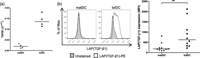
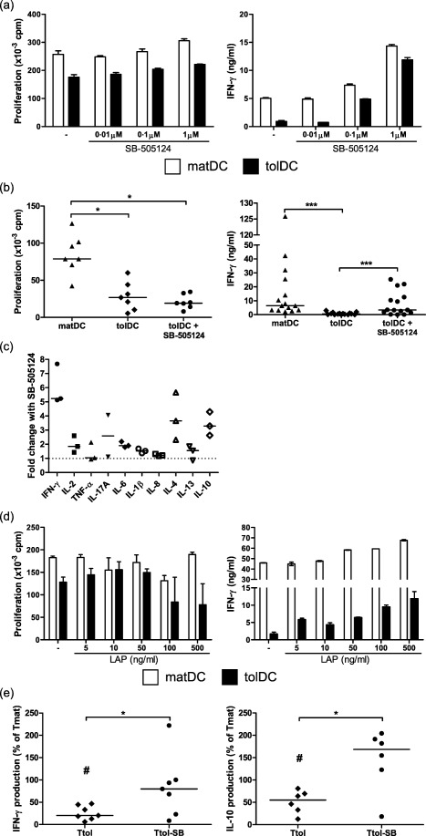
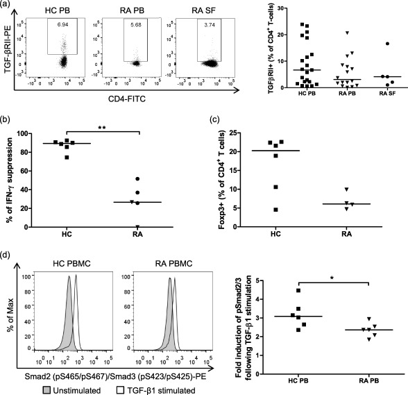
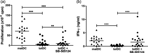
Tolerogenic dendritic cells (tolDC) are a new immunotherapeutic tool for the treatment of rheumatoid arthritis (RA) and other autoimmune disorders. We have established a method to generate stable tolDC by pharmacological modulation of human monocyte-derived DC. These tolDC exert potent pro-tolerogenic actions on CD4+ T cells. Lack of interleukin (IL)-12p70 production is a key immunoregulatory attribute of tolDC but does not explain their action fully. Here we show that tolDC express transforming growth factor (TGF)-β1 at both mRNA and protein levels, and that expression of this immunoregulatory cytokine is significantly higher in tolDC than in mature monocyte-derived DC. By inhibiting TGF-β1 signalling we demonstrate that tolDC regulate CD4+ T cell responses in a manner that is at least partly dependent upon this cytokine. Crucially, we also show that while there is no significant difference in expression of TGF-βRII on CD4+ T cells from RA patients and healthy controls, RA patient CD4+ T cells are measurably less responsive to TGF-β1 than healthy control CD4+ T cells [reduced TGF-β-induced mothers against decapentaplegic homologue (Smad)2/3 phosphorylation, forkhead box protein 3 (FoxP3) expression and suppression of (IFN)-γ secretion]. However, CD4+ T cells from RA patients can, nonetheless, be regulated efficiently by tolDC in a TGF-β1-dependent manner. This work is important for the design and development of future studies investigating the potential use of tolDC as a novel immunotherapy for the treatment of RA.

Keywords: TGF-β1; regulation; rheumatoid arthritis; tolerogenic dendritic cells.


Figures

Figure 1
Figure 1
Tolerogenic dendritic cells (tolDC) express transforming growth factor (TGF)‐β. (a) Expression of the TGF‐β1 gene by mature lipopolysaccharide (LPS)‐activated dendritic cells (matDC) and tolDC was measured using a custom Micro Fluidic Card (Applied Biosystems). mRNA expression was normalized to that of human glyceraldehyde 3‐phosphate dehydrogenase (GAPDH) by subtracting the CT value of the human GAPDH gene from the CT value of the gene of interest (ΔCT). Results are expressed as 2–ΔCT for four independent experiments. Horizontal lines represent median values. (b) matDC and tolDC were stained with anti‐human latency‐associated peptide (LAP) (TGF‐β1) antibody and were assessed by flow cytometry. Debris and dead cells were excluded on the basis of forward‐ and side‐scatter. Left panel shows one representative plot of 10 independent donors. The right panel shows the mean fluorescence intensity (MFI) for LAP (TGF‐β1) expression. Horizontal lines represent median values; n = 10. **P < 0·01 calculated with Wilcoxon signed‐rank test.
Figure 2
Figure 2
Transforming growth factor (TGF)‐β1 diminishes the ability of tolerogenic dendritic cells (tolDC) to stimulate healthy control (HC) T cells and is involved in their regulatory function. (a–d) Mature lipopolysaccharide (LPS)‐activated dendritic cells (matDC) or tolDC (1 × 10⁴ cells/well) were co‐cultured with allogeneic HC CD4+ T cells (1 × 10⁵ cells/well) in the absence or presence of increasing concentrations of SB‐505124, a small molecule inhibitor of TGF‐βRI (a) or 1 μM SB‐505124 (b,c) or increasing concentrations of recombinant latency‐associated peptide (LAP) (d). Proliferation (a,b,d, left panels), measured using [3H]‐thymidine uptake, and interferon (IFN)‐γ production (a,b,d, right panels), measured using enzyme‐linked immunosorbent assay (ELISA), were assessed at day 6. (c) Additional cytokines were detected in day 6 culture supernatants from tolDC‐CD4+ T‐cell cultures using an ELISA [interleukin (IL)−17A] or Meso Scale Discovery (MSD) immunoassay (all others). The fold change in cytokine production following inhibition of TGF‐β1 signalling was calculated by: concentration of cytokine produced in the presence of 1μM SB‐505124 ÷ concentration of cytokine produced in the absence of 1 μM SB‐505124. IL‐17A was undetectable in one experiment. Error bars in (a) and (d) represent standard error of the mean (s.e.m.) of triplicates (proliferation) or duplicates (IFN‐γ production). Horizontal lines in (b) and (c) represent median values, (b) left panel (proliferation) n = 7; (b) right panel (IFN‐γ production) n = 15; (c) (cytokines) n = 3. (e) Allogeneic healthy control (HC) CD4+ T cells were primed with DC (10 : 1) for 6 days and rested for 4 days with 10 IU/ml IL‐2. T cell lines primed by matDC (Tmat), tolDC (Ttol) and tolDC + SB‐505124 (Ttol‐SB) were restimulated with matDC and IFN‐γ (left panel) and IL‐10 (right panel) production, measured using ELISA, was assessed on day 3. Results are depicted as the percentage cytokine production of Tmat cell lines. Cytokine concentrations range: IFN‐γ in Ttol = 0.9–20·6 ng/ml and in Ttol‐SB = 4·4–80·7 ng/ml; IL‐10 in Ttol = 0·4–5·1 ng/ml and in Ttol‐SB = 0·9–31·7 ng/ml. Horizontal lines represent median values, n (IFN‐γ production) = 7; n (IL‐10 production) = 6. *P < 0·05 and ***P < 0·0001 calculated with Wilcoxon signed‐rank test. #Significant differences (P < 0·05) between Ttol and Tmat cells.
Figure 3
Figure 3
Healthy control (HC) and rheumatoid arthritis (RA) CD4+ T cells express transforming growth factor (TGF)‐βRII but RA CD4+ T cells have a reduced response to TGF‐β1 stimulation. Peripheral blood monuclear cells (PBMC) or CD4+ T cells were isolated from the peripheral blood (PB) of HC or RA patients and synovial fluid mononuclear cells (SFMC) were isolated from SF of RA patients. (a) Cells were stained with anti‐human CD3, anti‐human CD4 and anti‐human TGF‐βRII antibodies and were assessed by flow cytometry. Debris and dead cells were excluded on the basis of forward‐ and side‐scatter. A CD3+CD4+ gate was used to identify CD4+ T cells. Representative plots of TGF‐βRII expression on CD4+ T cells (left panel). The gate shows the percentage of CD4+ T cells expressing TGF‐βRII. The percentage of CD4+ T cells expressing TGF‐βRII (right panel). Horizontal lines represent median values; n (HC PB) = 21, n (RA PB) = 16, n (RA SF) = 5. (b) PBMC from HC (n = 6) and PBMC (▼) and SFMC (•) from RA patients (n = 5) were stimulated with αCD3αCD28 beads in the absence or presence of 10 ng/ml TGF‐β1 for 3 days. IFN‐γ was measured by enzyme‐linked immunosorbent assay (ELISA) and percentage suppression was calculated as follows: [(cytokine in absence of TGF‐β – cytokine in presence of TGF‐β)/cytokine in absence of TGF‐β × 100]. IFN‐γ concentrations range in cultures without TGF‐β1: HC = 1·1–32·4 ng/ml and RA patients = 0·7–32·2 ng/ml; and in cultures with TGF‐β1: HC = 0·1–5·3 ng/ml and RA patients = 0·5–79·1 ng/ml. (c) CD4+ T cells from HC PB (n = 6) and RA patient PB (n = 4) were stimulated with αCD3αCD28 beads in the presence of 10 ng/ml TGF‐β1 for 3 days. The percentage of CD4+forkhead box protein 3 (FoxP3)+ was determined by flow cytometry. (d) PBMC were left unstimulated or were stimulated with 10 ng/ml TGF‐β1 for 30 min before being fixed and permeabilized. PBMC were blocked with 2% mouse serum and stained with anti‐human CD3, anti‐human CD4 and anti‐human phospho mothers against decapentaplegic homologue (pSmad)2/3 antibodies before assessment by flow cytometry. Debris and dead cells were excluded on the basis of forward‐ and side‐scatter. A CD3+CD4+ gate was used to identify CD4+ T cells. Representative plots of pSmad2/3 expression on CD4+ T cells in unstimulated and stimulated conditions (left panel). Fold induction of pSmad2/3 in CD4+ T cells following TGF‐β1 stimulation calculated by dividing the mean fluorescence intensity (MFI) of stimulated cells by the MFI of unstimulated cells (right panel). Horizontal lines represent median values; n = 6. *P < 0·05 and **P < 0·01 calculated using a Mann‐Whitney U test.
Figure 4
Figure 4
Rheumatoid arthritis (RA) CD4+ T cells can be regulated by tolerogenic dendritic cells (tolDC) in a transforming growth factor (TGF)‐β‐dependent manner. CD4+ T cells (1 × 10⁵ cells/well) from RA patients were co‐cultured with allogenic mature lipopolysaccharide (LPS)‐activated dendritic cells (matDC) or tolDC (1 × 10⁴ cells/well) from a healthy control in the absence or presence of 1 μM SB‐505124, a small molecule inhibitor of TGF‐βRI. Proliferation (left panel), measured using [3H]‐thymidine uptake, and interferon (IFN)‐γ production (right panel), measured using enzyme‐linked immunosorbent assay (ELISA), were assessed at day 6; n = 18. Horizontal lines represent median values. **P < 0·01 and ***P < 0·0005 calculated with Wilcoxon signed‐rank test.

Similar articles

Cited by

References

    1. Boissier MC, Semerano L, Challal S, Saidenberg‐Kermanac'h N, Falgarone G. Rheumatoid arthritis: from autoimmunity to synovitis and joint destruction. J Autoimmun 2012; 39:222–8. - PubMed
    1. Anderson AE, Sayers BL, Haniffa MA et al Differential regulation of naive and memory CD4+ T cells by alternatively activated dendritic cells. J Leukoc Biol 2008; 84:124–33. - PMC - PubMed
    1. Anderson AE, Swan DJ, Sayers BL et al LPS activation is required for migratory activity and antigen presentation by tolerogenic dendritic cells. J Leukoc Biol 2009; 85:243–50. - PMC - PubMed
    1. Harry RA, Anderson AE, Isaacs JD, Hilkens CM. Generation and characterisation of therapeutic tolerogenic dendritic cells for rheumatoid arthritis. Ann Rheum Dis 2010; 69:2042–50. - PMC - PubMed
    1. Stoop JN, Harry RA, von Delwig A, Isaacs JD, Robinson JH, Hilkens CM. Therapeutic effect of tolerogenic dendritic cells in established collagen‐induced arthritis is associated with a reduction in Th17 responses. Arthritis Rheum 2010; 62:3656–65. - PubMed

MeSH terms