. 2015 Jul 15;2(11):1500122.
doi: 10.1002/advs.201500122. eCollection 2015 Nov.

Injectable Hydrogels for Cardiac Tissue Repair after Myocardial Infarction

Affiliations

Injectable Hydrogels for Cardiac Tissue Repair after Myocardial Infarction

Anwarul Hasan et al. Adv Sci (Weinh). .

Abstract

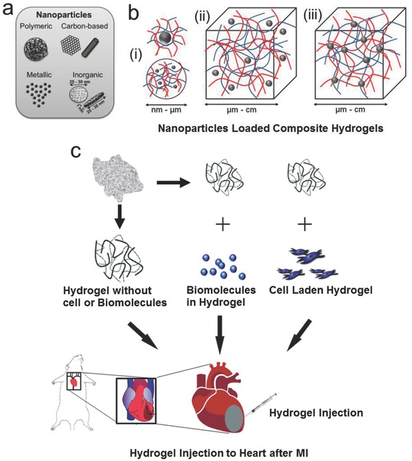
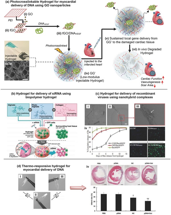
Cardiac tissue damage due to myocardial infarction (MI) is one of the leading causes of mortality worldwide. The available treatments of MI include pharmaceutical therapy, medical device implants, and organ transplants, all of which have severe limitations including high invasiveness, scarcity of donor organs, thrombosis or stenosis of devices, immune rejection, and prolonged hospitalization time. Injectable hydrogels have emerged as a promising solution for in situ cardiac tissue repair in infarcted hearts after MI. In this review, an overview of various natural and synthetic hydrogels for potential application as injectable hydrogels in cardiac tissue repair and regeneration is presented. The review starts with brief discussions about the pathology of MI, its current clinical treatments and their limitations, and the emergence of injectable hydrogels as a potential solution for post MI cardiac regeneration. It then summarizes various hydrogels, their compositions, structures and properties for potential application in post MI cardiac repair, and recent advancements in the application of injectable hydrogels in treatment of MI. Finally, the current challenges associated with the clinical application of injectable hydrogels to MI and their potential solutions are discussed to help guide the future research on injectable hydrogels for translational therapeutic applications in regeneration of cardiac tissue after MI.

Keywords: cardiac repair; hydrogels; myocardial infarction; regenerative medicine; stem cell; tissue engineering.


Figures

Figure 1
Figure 1
Chemical structures of various natural and synthetic hydrogels that have been tried or have the potential for application as injectable hydrogels in cardiac tissue regeneration. a) Some natural hydrogels: i) collagen,134 ii) gelatine,135 iii) hyaluronic acid,135 iv) alginate,136 v) agarose,137 vi) chitosan,138 and vii) keratin. b) Some synthetic hydrogels: i) polyacrylic acid, ii) poly(ethylene oxide), iii) polyvinyl alcohol, iv) polyphosphazene, v) polypeptide chains.
Figure 2
Figure 2
Schematic representation of nanoparticle loaded injectable composite hydrogels, and delivery of various hydrogels including acellular hydrogels alone, acellular hydrogels with various biomolecules and hydrogels with various cells. a) A range of nanoparticles such as polymeric nanoparticles, metallic‐metal oxide based nanoparticles, inorganic nanoparticles, and carbon‐based nanomaterials can be incorporated in hydrogels to make nanocomposite hydrogels. Adapted with permission.109 b) The common strategies for incorporation of nanoparticles in hydrogels: i) stabilization of inorganic or polymeric nanoparticles by nano–micro‐sized hydrogel particles, ii) noncovalently immobilized nanoparticles in a hydrogel matrix, and iii) covalently immobilized nanoparticles in hydrogel matrix. Adapted with permission.110 c) Different strategies of injectable hydrogel delivery for treatment of MI. The injectable hydrogels can be delivered alone without any biomolecules or cells, with some biomolecules as carriers or with cells as a 3D matrix. Adapted with permission.76 Copyright 2010, Royal Society of Chemistry.
Figure 3
Figure 3
a–h) Enhancement of cardiac muscle and reduction of infarct fibrosis using myocardial matrix. Histological characterization of infarcted pig hearts: Masson's trichrome staining images are representative of six matrix‐injected pigs and four control animals. a) Matrix‐injected hearts had a distinct, thick endocardium (red stained muscle, indicated with an asterisk). b) Noninjected control animals had a loose fibrillar layer (blue) beneath the endothelium. c) In saline injected control animals, the endocardium was moderately thickened with minimal muscle (red). d) An adjacent tissue section for the matrix‐injected animal in a) stained for cardiac troponin‐T, indicating the presence of cardiomyocytes. e) Area of endocardial layer of muscle as a proportion of the infarct. f) Percentage of collagen content in the infarcts. Data are means ± SEM and were obtained from Masson's trichrome slides a–c). *P < 0.05 (Student's t test). g,h) Matrix‐injected hearts contained foci of neovascularization in the area below the endocardium (g, arrows), but none of the saline or noninjected control hearts showed these areas of neovascularization h). Scale bar, 200 mm. i) Myocardial matrix is biocompatible and biodegradable. Representative histological sections of cell infiltration in matrix‐injected rat hearts injected with saline, PMM, or NDM at days 3, 14, and 28. Inflammation and multinucleated giant cells are present in the NDM groups at days 14 and 28 (arrows). The PMM (asterisk marked porous network) was completely degraded by 28 d. Scale bar, 200 mm. Reproduced with permission.78 Copyright 2013, American Association for the Advancement of Science.
Figure 4
Figure 4
Arterial staining and histological morphology of excised rat hearts treated with injected hydrogels 4 weeks post MI. Image (a) shows Alpha‐SMA positive vessels in the infarct and peri‐infarct zones, 4 weeks after MI. Panels (i) and (ii) show control (saline injection) peri‐infarct and infarct zone respectively, (iii) and (iv) show peri‐infarct and infarct treated with PF1%, (v) and (vi) show those treated with PF2% respectively, (vii) and (viii) treated with TF1%, respectively, and (ix) and (x) treated with TF2%, respectively. In image (b) 4 weeks after the MI the treated rat hearts were cut into equal transverse slices to show the gross appearance and histological morphology (i) in that of the untreated saline control group where saline injection was only used. The PF1% hydrogel injection in (ii), the PF2% hydrogel injection in (iii), the TF1% hydrogel injection in (iv), and the TF2% hydrogel injection in (v). Reproduced with permission.13 Copyright 2014, Elsevier. Image (c) shows immune‐histochemical staining for the smooth muscles for the four groups 30 d after MI. Panel (i) shows the control group, (ii) shows hydrogel group, (iii) shows BMMNC group, and (iv) shows BMMNC + hydrogel group. Reproduced with permission.124 Image (d) shows the staining with Masson trichrome of the infarcted wall forcollagen (green) and muscle (red) of the four groups. (i) PBS‐only group of the four groups; (ii) OPF‐only group; (iii) PBS + ESC group; and (iv) OPF + ESC group. Reproduced with permission.19 Copyright 2014, Elsevier. Image (e) also shows staining with Masson's trichrome of the infarct area and shows similar level of tissue improvement with BADSCs treatment alone or Chitosan alone compared to the control group with Chitosan hydrogel delivery of BADSCs resulting in the greatest healing and infarct reduction. Reproduced with permission.124
Figure 5
Figure 5
a–d) Structural, physical, and electrical properties of CNT‐GelMA hydrogels: a) schematic diagram illustrating the isolated heart conduction systems showing the purkinje fibers, which are located in the inner ventricular walls of the heart. b) Preparation procedure of fractal‐like CNT networks embedded in GelMA hydrogel. c) TEM image of GelMA‐coated CNTs. d) SEM images show porous surfaces of a 1 mg mL−1 CNT‐GelMA thin film. e,f) Adhesion, maturation, alignment, and phenotype of cardiac cells on CNT‐GelMA hydrogels. e) Confocal images of cardiomyocytes after culturing for 5 d on pristine GelMA and 1 mg mL−1 CNT‐GelMA revealed more uniform cell distribution and partial cell alignment on CNT‐GelMA. Higher magnification images showed well‐elongated cardiac cells and well‐developed F‐actin cross‐striations (bottom right, white arrows) on CNT‐GelMA but not on pristine GelMA (bottom left). f) Immunostaining of sarcomeric R‐actinin (green), nuclei (blue), and Cx‐43 (red) revealed that cardiac tissues (8 d culture) on (i) pristine GelMA and (ii) CNT GelMA were phenotypically different. Partial uniaxial sarcomere alignment and interconnected sarcomeric structure with robust intercellular junctions were observed on CNT‐GelMA. Immunostaining of Troponin I (green) and nuclei (blue) showed much less and more aggregated Troponin I presence on (iii) pristine GelMA than on (iv) CNT‐GelMA. g–i) Improved mechanical integrity and advanced electrophysiological functions of cardiac tissues on CNT‐GelMA. g) Spontaneous beating rates of cardiac tissues recorded from day 3 to day 9 on a daily basis. h) CNTs protected cardiac tissues against damages by heptanol. Plots of spontaneous beating amplitude over time (5 d culture) for 0–5 mg mL−1 CNTs in GelMA in response to 4 × 10⁻³ m heptanol. i) Time lapse before sporadic beating and stop of beating induced by heptanol (*p < 0.05).Reproduced with permission.125 Copyright 2013, American Chemical Society.
Figure 6
Figure 6
Injectable hydrogels for gene delivery applications. a) Photocrosslinkable hydrogel for myocardial delivery of vascular endothelial growth factor (VEGF) carrying gene using cationic functionalized graphene oxide (fGO) nanoparticles. Schematic of stepwise formulation process for direct intramyocardial injection of damaged heart with acute myocardial infarction. Reproduced with permission.126 Copyright 2014, American Chemical Society. b) Delivery of siRNA using biopolymer hydrogel—schematic of hydrogel formation for delivery of siRNA and subsequent inhibition of gene expression in incorporated and neighboring cells. Biomaterial solutions of alginate, photo alginate, or collagen are mixed with siRNA and GFP‐positive cells, and hydrogels are then formed by crosslinking, photo‐crosslinking, or thermos‐gelling, respectively. The siRNA diffuses through the hydrogel to affect incorporated cells, and it is also released from the hydrogel to locally affect surrounding cells that are part of the host tissue. Reproduced with permission.127 Copyright 2009, American Chemical Society. c) Hydrogel for delivery of recombinant viruses using nanohybrid complexes. Controlled release of baculovirus (Bac) using CNT reinforced hydrogel. TEM images of (i) nonfunctionalized CNT with Bac, (ii) CNT functionalized Bac with arrow showing the baculovirus bound to CNT surface in magnified image (iii). Scale bar indicates 100 nm length. Self‐assembled nanocomplex of cationic PAA functionalized CNTs (f‐CNT) were hybridized with anionic Bac. (iii) Cumulative release kinetics of baculovirus from denatured collagen hydrogel (2.5 mg mL−1) impregnated with Bac–CNT nanocomplex (v–viii–G) rMSCs were overlaid on the collagen hydrogel formulated with CNT (25 μg mL−1), BacMGFP, CNT/BacMGFP or functionalized CNT f‐CNT/BacMGFP. Abbreviations: TEM = transmission electron microscope; CNT = carbon nanotube; rMSCs = rat mesenchymal stem cells; PAA = poly (acrylic acid), MGFP = Monster Green Fluorescent Protein. Reproduced with permission.128 Copyright 2014, Elsevier. d) Thermo‐responsive hydrogel for myocardial delivery of plasmid DNA. Thermosensitive sol‐to‐gel transition properties of biodegradable dextran‐poly(e‐caprolactone)‐2‐hydroxylethylmethacrylate‐poly(N‐isopropylacryl amide) (Dex‐PCL‐HEMA/PNIPAAm) hydrogel at 1.5 wt% concentration: (i) gel solution in fluidity at room temperature, (ii) the gel solution turned into gel at 37 °C, and (iii) sol–gel reversibility of gel solution at room temperature. (iv) Representative pictures of left ventricles from each group after Masson's Trichrome staining 30 d after treatments. Scale bar = 1 mm. Below images are the infarct size as percentages at 30 d. Infarct size is calculated as the ratio of infarcted to noninfarcted area of the left ventricle. Reproduced with permission.129

References

    1. Curtis M. W., Russell B., J. Cardiovasc. Nurs. 2009, 24, 87. - PMC - PubMed
    1. Radhakrishnan J., Krishnan U. M., Sethuraman S., Biotechnol. Adv. 2014, 32, 449. - PubMed
    1. Slaughter B. V., Khurshid S. S., Fisher O. Z., Khademhosseini A., Peppas N. A., Adv. Mater. 2009, 21, 3307. - PMC - PubMed
    1. Lee K. Y., Mooney D. J., Chem. Rev. 2001, 101, 1869. - PubMed
    1. Kolettis T. M., Vilaeti A., Dimos K., Tsitou N., Agathopoulos S., Mini Rev. Med. Chem. 2011, 11, 263. - PubMed